

doi: 10.1002/anie.200901668.
Three-dimensional nanostructured substrates toward efficient capture of circulating tumor cells
Affiliations
- PMID: 19847834
- PMCID: PMC2878179
- DOI: 10.1002/anie.200901668
Item in Clipboard
Three-dimensional nanostructured substrates toward efficient capture of circulating tumor cells
Angew Chem Int Ed Engl.
2009.
No abstract available
Figures
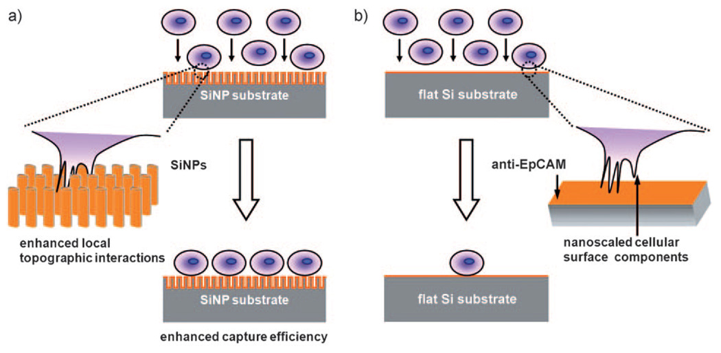
Conceptual illustration of how an anti-EpCAM-coated 3D nanostructured (i.e., SiNP) substrate can be employed to achieve significantly enhanced capture of EpCAM-positive cells (i.e., CTCs) from cell suspension in contrast to an anti-EpCAM-coated unstructured (i.e., flat Si) substrate. a) Interdigitation of nanoscale cellular surface components and SiNPs enhances local topographic interactions, resulting in vastly improved cell-capture efficiency. b) Lack of local topographic interactions between cells and flat Si substrate compromises the respective cell-capture efficiency.
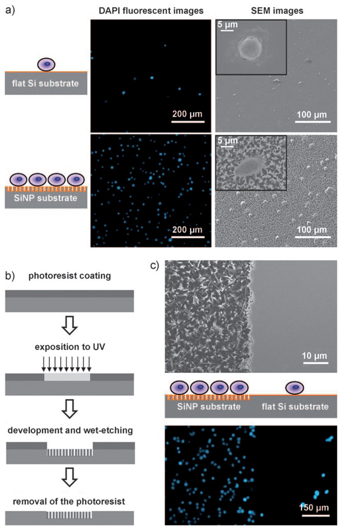
a) Fluorescence micrographs and SEM images of SiNP substrates and flat Si substrates on which MCF7 cells were captured. The SiNP substrates exhibited significantly better cell-capture efficiency than the flat ones. DAPI: 4’,6-Diamidino-2-phenylindol. b) Photolithography process for patterning alternate SiNP and flat substrates on the silicon wafer for comparing their cell-capture efficiencies in a close experimental setup. c) SEM images of patterned and flat substrates before cell capture (top) and fluorescence micrographs of cells captured on patterned and flat substrates (bottom).

Quantitative evaluations of cell-capture yields a) at different capture times and b) with different SiNP lengths ranging from 0 to 20 mm. Each plot and error bar represents a mean standard deviation from three repeats.

A) Chemical etching by Ag+ and HF was employed to produce a silicon nanopillar (SiNP) array on a silicon wafer. The SEM images reveal that well-defined SiNPs with diameters ranging from 100 to 200 nm and lengths around 10 µm were produced. B) Grafting of biotinylated epithelial-cell adhesion-molecule antibody (anti-EpCAM) onto silicon substrates.
References
-
- Steeg PS. Nat. Med. 2006;12:895. - PubMed
-
- Budd GT, Cristofanilli M, Ellis MJ, Stopeck A, Borden E, Miller MC, Matera J, Repollet M, Doyle GV, Terstappen LWMM, Hayes DF. Clin. Cancer Res. 2006;12:6403. - PubMed
-
- Allard WJ, Matera J, Miller MC, Repollet M, Connelly MC, Rao C, Tibbe AGJ, Uhr JW, Terstappen LWMM. Clin. Cancer Res. 2004;10:6897. - PubMed
-
- Zieglschmid V, Hollmann C, Böcher O. Crit. Rev. Clin. Lab. Sci. 2005;42:155. - PubMed
-
- Pantel K, Brakenhoff RH, Brandt B. Nat. Rev. Cancer. 2008;8:329. - PubMed
Publication types
MeSH terms
Substances
Grants and funding
LinkOut - more resources
Full Text Sources
Other Literature Sources
