. 2009 Oct;32(10):1313-24.
doi: 10.1093/sleep/32.10.1313.

Modulation of genioglossus muscle activity across sleep-wake states by histamine at the hypoglossal motor pool

Affiliations

Modulation of genioglossus muscle activity across sleep-wake states by histamine at the hypoglossal motor pool

Timothy Bastedo et al. Sleep. 2009 Oct.

Abstract

Study objectives: Histamine neurons comprise a major component of the aminergic arousal system and significantly influence sleep-wake states, with antihistamines widely used as sedative hypnotics. Unlike the serotonergic and noradrenergic components of this arousal system, however, the role of histamine in the central control of respiratory motor activity has not been determined. The aims of this study were to characterize the effects of histamine receptor agonists and antagonists at the hypoglossal motor pool on genioglossus muscle activity across sleep and awake states, and also determine if histamine contributes an endogenous excitatory drive to modulate hypoglossal motor outflow to genioglossus muscle.

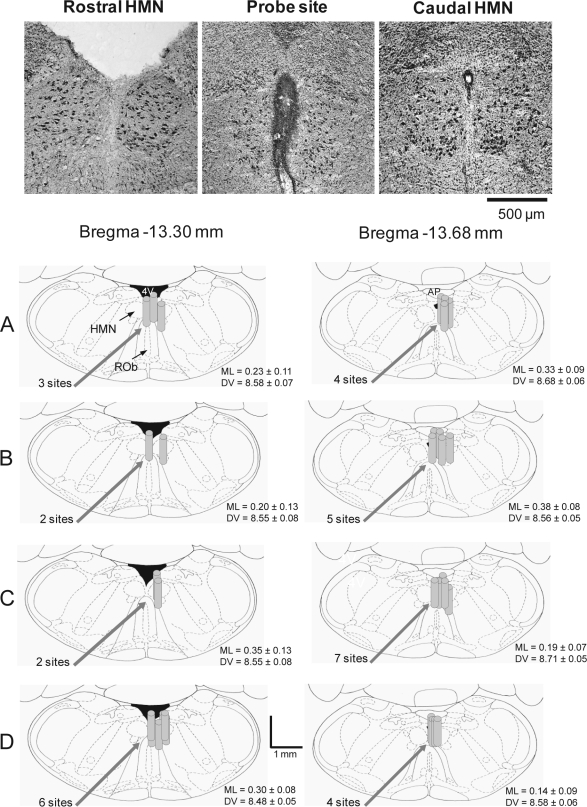
Design, participants, and interventions: Thirty-three rats were implanted with electroencephalogram and neck electrodes to record sleep-wake states, and genioglossus and diaphragm electrodes for respiratory muscle recordings. Microdialysis probes were inserted into the hypoglossal motor nucleus.

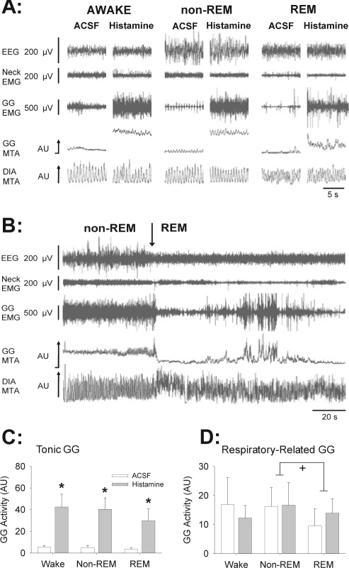
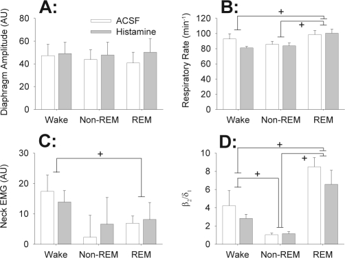
Measurements and results: Histamine at the hypoglossal motor nucleus significantly increased tonic genioglossus muscle activity in wakefulness, non-REM sleep and REM sleep. The activating effects of histamine on genioglossus muscle activity also occurred with a histamine type-1 (H1) but not H2 receptor agonist. However, H1 receptor antagonism at the hypoglossal motor nucleus did not decrease genioglossus muscle activity in wakefulness or sleep.

Conclusions: The results suggest that histamine at the hypoglossal motor pool increases genioglossus muscle activity in freely behaving rats in wakefulness, non-REM, and REM sleep via an H1 receptor mechanism.


Figures

Figure 1
Figure 1
Example and group data showing location of the microdialysis probes from all the experiments. The top images show histological sections with a lesion site made by the microdialysis probe immediately adjacent to both hypoglossal motor nuclei. Also shown are coronal diagrams from the rat medulla, illustrating the distribution of individual microdialysis sites from all rats administered (A) histamine, (B) the H1 receptor agonist 2-(2-pyridyl)ethylamine, (C) the H2 receptor agonist dimaprit, and (D) the H1 receptor antagonist diphenhydramine. The microdialysis probe locations are represented by gray cylinders which are drawn to scale. Overlap obscures some of the dialysis sites. The mean stereotaxic coordinates for the center of the probe sites are given in the medial-lateral (ML) and dorsal-ventral (DV) directions for each study and for each plane posterior to bregma. Abbreviations: AP, area postrema; HMN, hypoglossal motor nucleus; 4V, fourth ventricle.
Figure 2
Figure 2
Stimulating effects of histamine at the hypoglossal motor pool on genioglossus (GG) muscle activity across sleep-wake states. (A) Traces show the electroencephalogram (EEG), neck electromyogram (EMG), GG, and diaphragm (DIA) EMG signals. The GG and DIA signals are also displayed as their moving-time averages (MTA) in arbitrary units (AU). The baseline of the integrator (i.e., electrical zero) is shown for the GG MTA. Compared to artificial cerebrospinal fluid (ACSF) controls, histamine increased tonic GG activity across all sleep-wake states. (B) Traces show that despite the persistence of tonic GG activation in REM sleep with histamine, GG activity was still decreased in REM compared to levels observed in the preceding period of non-REM sleep. (C and D) Group data showing a significant excitatory effect of histamine on tonic GG activity in wakefulness, non-REM, and REM sleep (C); but with no effects on respiratory related GG activity (D). The symbols * and + indicate P < 0.05 compared to ACSF controls, and between the indicated sleep-wake states, respectively. The suppression of respiratory-related GG activity in REM sleep (panel D) was independent of whether ACSF or histamine was applied to the hypoglossal motor pool. All data are shown as mean + SEM (n = 7 rats). The mean values for each individual rat were first calculated from the population of values that occurred for all the 5-sec epochs during each sleep-wake state with each drug applied to the hypoglossal motor pool. The means from each individual rat in each condition were then averaged to yield the grand means for the group which are shown in the figure. See text for further details.
Figure 3
Figure 3
Responses to histamine at the hypoglossal motor pool were specific to the genioglossus muscle as there were no significant effects on (A) diaphragm amplitude, (B) respiratory rate, (C) neck muscle activity and (D) the ratio of high (20–30 Hz) to low (0.5–2 Hz) frequency activity in the electroencephalogram (i.e., the β21 ratio). All data are shown as mean + SEM (n = 7 rats). Data were averaged as described in Figure 2 and the Methods. The symbol + indicates P < 0.05 between the indicated sleep-wake states. The changes in these variables were independent of whether ACSF or histamine was applied to the hypoglossal motor pool. See text for further details.
Figure 4
Figure 4
Similar to the effects of histamine, H1 receptor agonism at the hypoglossal motor pool also increased GG activity across sleep-wake states. (A) Example traces showing that compared to ACSF controls, application of the H1 receptor agonist 2-(2-pyridyl)ethylamine (PEA), increased tonic GG activity across sleep-wake states. (B) Group data showing a significant stimulating effect of PEA on tonic and respiratory-related GG activities in wakefulness, non-REM, and REM sleep. The symbols * and + indicate P < 0.05 compared to ACSF controls, and between the indicated sleep-wake states, respectively. The suppression of tonic GG activity in REM sleep, compared to wakefulness and non-REM sleep (panel B), occurred with 1mM PEA only. See text for further details. All data are shown as mean + SEM (n = 7 rats). Data were averaged as described in Figure 2 and the Methods. Abbreviations are as for Figure 2.
Figure 5
Figure 5
Example traces (A) and group data (B) showing that H2 receptor agonism at the hypoglossal motor pool did not significantly alter GG activity. + indicates P < 0.05 between the indicated sleep-wake states, with the suppression of respiratory-related GG activity in REM sleep (panel C) being independent of whether ACSF or 0.1 mM dimaprit was applied to the hypoglossal motor pool. All data are shown as mean + SEM. Six rats were studied with ACSF and 0.1 mM dimaprit, and 6 were studied with ACSF and 1 mM dimaprit, with 3 rats receiving both doses. Data were averaged as described in Figure 2 and the Methods. Abbreviations are as for Figure 2. See text for further details.
Figure 6
Figure 6
Group data showing that H1 receptor antagonism at the hypoglossal motor pool did not significantly alter tonic (A) or respiratory-related (B) GG activity, indicating a minimal endogenously active H1 receptor mediated excitatory drive in these freely behaving animals across natural sleep-wake states. All data are shown as mean + SEM (n = 10 rats). Data were averaged as described in Figure 2 and the Methods.

Similar articles

Cited by

References

    1. Jones BE. Basic mechanisms of sleep-wake states. In: Kryger MH, Roth T, Dement WC, editors. Principles and practice of sleep medicine. 2nd ed. Philadelphia: Saunders; 1994. pp. 145–62.
    1. Jacobs BL, Azmitia EC. Structure and function of the brain serotonin system. Physiol Rev. 1992;72:165–229. - PubMed
    1. Foote SL, Bloom FE, Aston-Jones G. Nucleus locus ceruleus: new evidence of anatomical and physiological specificity. Physiol Rev. 1983;63:844–914. - PubMed
    1. Saper CB, Scammell TE, Lu J. Hypothalamic regulation of sleep and circadian rhythms. Nature. 2005;437:1257–63. - PubMed
    1. Rekling JC, Funk GD, Bayliss DA, Dong XW, Feldman JL. Synaptic control of motoneuronal excitability. Physiol Rev. 2000;80:767–852. - PMC - PubMed

Publication types

MeSH terms