. 2010 Jan 1;468(1):12-7.
doi: 10.1016/j.neulet.2009.10.047. Epub 2009 Oct 20.

Comparison of melanin-concentrating hormone and hypocretin/orexin peptide expression patterns in a current parceling scheme of the lateral hypothalamic zone

Affiliations

Comparison of melanin-concentrating hormone and hypocretin/orexin peptide expression patterns in a current parceling scheme of the lateral hypothalamic zone

Joel D Hahn. Neurosci Lett. .

Abstract

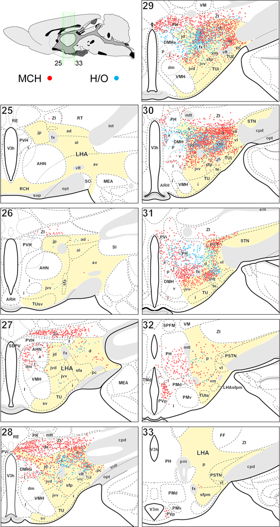
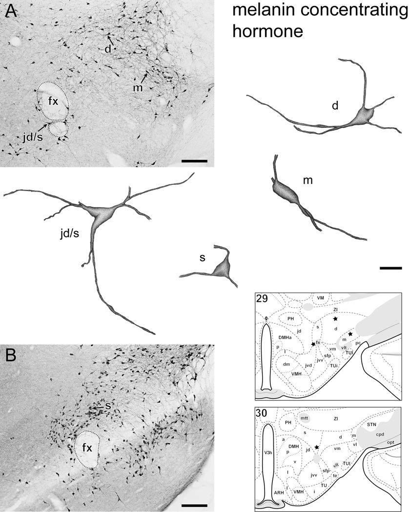
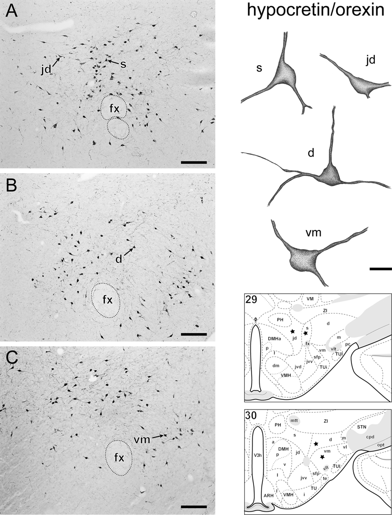
The distribution of hypothalamic neurons expressing the peptides melanin-concentrating hormone (MCH; 'MCH neurons') or hypocretin/orexin (H/O; 'H/O neurons') was assessed with immunocytochemistry in male rats at high spatial resolution. Data were plotted on a rat brain atlas that includes a recently revised parcellation scheme for the lateral hypothalamic zone. Quantitative analysis revealed approximately three times more MCH neurons than H/O neurons in the hypothalamus, and approximately twice as many within the parcellations of the lateral hypothalamic area (LHA). The LHA contained 60% of MCH neurons and 81% of H/O neurons, and the same five LHA regions contained the vast majority of MCH (87%) or H/O (93%) neurons present within the LHA: namely the LHA dorsal region (LHAd: 31% of H/O; 38% of MCH), suprafornical region (LHAs: 28% of H/O; 11% of MCH), ventral region medial zone (LHAvm: 15% of H/O; 16% of MCH), juxtadorsomedial region (LHAjd: 14% of H/O and MCH) and magnocellular nucleus (LHAm: 5% of H/O; 7% of MCH). The zona incerta (ZI) contained 18% of MCH neurons. A high co-abundance of MCH and H/O neurons outside of the LHA was present in the posterior hypothalamic nucleus (PH: 11% of H/O; 9% of MCH). Morphological analysis revealed MCH and H/O neurons as typically tri-polar with irregularly shaped somata. These data provide a quantitative analysis of neurons expressing either MCH or H/O peptides within the rat hypothalamus, and they clarify differences in the distribution pattern for different subsets of these neuron types, especially within the LHA.


Figures

Figure 1
Figure 1
Distribution of MCH- and ppH/O-immunoreactive somata from 5 adult male rats plotted on a reference atlas of the rat hypothalamus; the lateral hypothalamic zone is colored yellow. Each colored dot represents a single MCH (red) or H/O (blue) neuron; the dots are semi-transparent such that overlapping MCH or H/O neurons are indicated by greater color saturation or shades of purple. The number of plotted cells equals that counted in 2 series (one each for MCH and one for H/O) from 5 series of 25-µm-thick transverse sections obtained from 5 different rats. Immunoreactive somata were mapped with camera lucida from adjacent series of sections and with reference to an additional series from the same rats stained with thionin. The diagram upper left indicates in midsagittal view the location and extent of the reference atlas levels on to which data were plotted. For lateral zone abbreviations see Table 1, other: AHN, anterior hypothalamic nucleus; ARH, arcuate nucleus; cpd, cerebral peduncle; DMHa,p,v, dorsomedial nucleus anterior, posterior, ventral parts; FF, fields of Forel; fx, fornix; I, internuclear area; int, internal capsule; MEA, medial amygdalar nucleus; mtt, mammillothalamic tract; opt, optic tract; PH, posterior hypothalamic nucleus; pm, principal mammillary tract; PMd,v, dorsal and ventral premammillary nuclei; PR, perireuniens nucleus; PVH paraventricular nucleus; PVHf, PVH forniceal part; PVi, intermediate periventricular nucleus; PVp, posterior periventricular nucleus; RE, nucleus reuniens; RT, reticular nucleus; SBPV, subparaventricular region; SI, substantia innominata; SO, supraoptic nucleus; sup, supraoptic commissures; TMd, dorsal tuberomammillary nucleus; V3h, third ventricle hypothalamic part; V3m, third ventricle mammillary recess; vlt, ventrolateral hypothalamic tract; VM, ventral medial nucleus; VMHdm, ventromedial nucleus dorsomedial part; ZI, zona incerta; ZIda, ZI dopaminergic group.
Figure 2
Figure 2
Examples of individual neurons immunoreactive for MCH (Fig. 2) or ppH/O (Fig. 3) within the LHA. 2A, B and 3A-C, photomicrographs showing MCH (2A, B) and ppH/O (3A-C) immunoreactive (ir) neurons within the lateral hypothalamic zone. The soma and dendrites of selected neurons within subregions of the LHA (identified with reference to corresponding thionin stained sections) were drawn with camera lucida. Individual neurons were traced as far as visible within 25-µm-thick transverse sections. Drawings are stippled and shaded to enhance the appearance of 3-dimensions. The location of each drawn neuron is indicated by an arrow on the photomicrograph with a corresponding letter(s) indicating LHA subdivision. For reference, approximate neuron location is shown on a corresponding rat brain atlas -- asterisks indicate approximate location of drawn neurons in each figure. Abbreviations: d, jd, m, s, vm, (respectively) LHA -dorsal region, -juxtadorsomedial region, -magnocellular nucleus, -suprafornical region, -ventral region medial zone; fx = fornix; for other atlas abbreviations see Fig. 1. Bars = 200 µm photomicrographs, 20 µm drawings.
Figure 3
Figure 3
Examples of individual neurons immunoreactive for MCH (Fig. 2) or ppH/O (Fig. 3) within the LHA. 2A, B and 3A-C, photomicrographs showing MCH (2A, B) and ppH/O (3A-C) immunoreactive (ir) neurons within the lateral hypothalamic zone. The soma and dendrites of selected neurons within subregions of the LHA (identified with reference to corresponding thionin stained sections) were drawn with camera lucida. Individual neurons were traced as far as visible within 25-µm-thick transverse sections. Drawings are stippled and shaded to enhance the appearance of 3-dimensions. The location of each drawn neuron is indicated by an arrow on the photomicrograph with a corresponding letter(s) indicating LHA subdivision. For reference, approximate neuron location is shown on a corresponding rat brain atlas -- asterisks indicate approximate location of drawn neurons in each figure. Abbreviations: d, jd, m, s, vm, (respectively) LHA -dorsal region, -juxtadorsomedial region, -magnocellular nucleus, -suprafornical region, -ventral region medial zone; fx = fornix; for other atlas abbreviations see Fig. 1. Bars = 200 µm photomicrographs, 20 µm drawings.

Similar articles

Cited by

References

    1. Goto M, Canteras NS, Burns G, Swanson LW. Projections from the subfornical region of the lateral hypothalamic area. J Comp Neurol. 2005;493:412–438. - PMC - PubMed
    1. Elias CF, Saper CB, Maratos-Flier E, Tritos NA, Lee C, Kelly J, Tatro JB, Hoffman GE, Ollmann MM, Barsh GS, Sakurai T, Yanagisawa M, Elmquist JK. Chemically defined projections linking the mediobasal hypothalamus and the lateral hypothalamic area. J. Comp Neurol. 1998;402:442–459. - PubMed
    1. Abrahamson EE, Moore RY. The posterior hypothalamic area: chemoarchitecture and afferent connections. Brain Res. 2001;889:1–22. - PubMed
    1. Bittencourt JC, Presse F, Arias C, Peto C, Vaughan J, Nahon JL, Vale W, Sawchenko PE. The melanin-concentrating hormone system of the rat brain: an immuno- and hybridization histochemical characterization. J Comp Neurol. 1992;319:218–245. - PubMed
    1. Cvetkovic V, Brischoux F, Jacquemard C, Fellmann D, Griffond B, Risold PY. Characterization of subpopulations of neurons producing melanin-concentrating hormone in the rat ventral diencephalon. J. Neurochem. 2004;91:911–919. - PubMed

Publication types

LinkOut - more resources