. 2010 Jan;43(1):136-45.
doi: 10.1016/j.mcn.2009.10.004. Epub 2009 Oct 20.

Astrocyte secreted proteins selectively increase hippocampal GABAergic axon length, branching, and synaptogenesis

Affiliations

Astrocyte secreted proteins selectively increase hippocampal GABAergic axon length, branching, and synaptogenesis

Ethan G Hughes et al. Mol Cell Neurosci. 2010 Jan.

Abstract

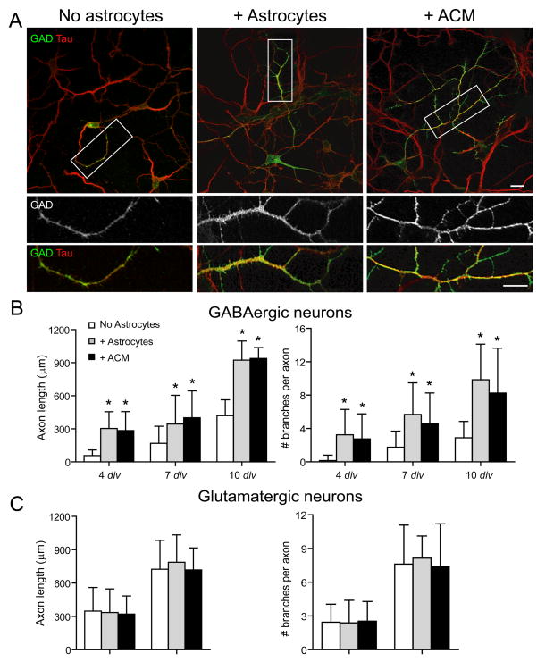
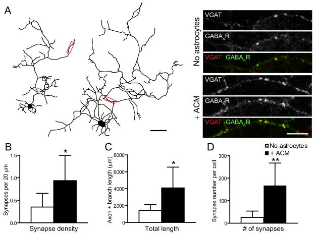
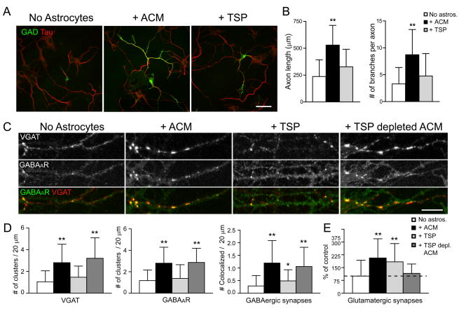
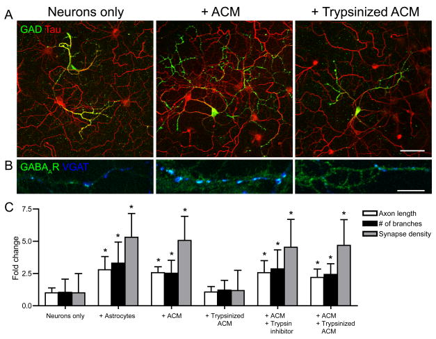
Astrocytes modulate the formation and function of glutamatergic synapses in the CNS, but whether astrocytes modulate GABAergic synaptogenesis is unknown. We demonstrate that media conditioned by astrocytes, but not other cells, enhanced GABAergic but not glutamatergic axon length and branching, and increased the number and density of presynaptically active GABAergic synapses in dissociated hippocampal cultures. Candidate mechanisms and factors, such as activity, neurotrophins, and cholesterol were excluded as mediating these effects. While thrombospondins secreted by astrocytes are necessary and sufficient to increase hippocampal glutamatergic synaptogenesis, they do not mediate astrocyte effects on GABAergic synaptogenesis. We show that the factors in astrocyte conditioned media that selectively affect GABAergic neurons are proteins. Taken together, our results show that astrocytes increase glutamatergic and GABAergic synaptogenesis via different mechanisms and release one or more proteins with the novel functions of increasing GABAergic axon length, branching and synaptogenesis.


Figures

Figure 1
Figure 1. Astrocytes selectively increase GABAergic axon length and branching
Hippocampal neurons were cultured alone, with astrocytes or ACM and were immunostained at 4, 7, and 10 div with antibodies against tau (red) and glutamic acid decarboxylase (GAD; green). (A), GABAergic axon length and branching were significantly increased in neurons cultured with astrocytes (middle) or ACM (right) compared to neurons cultured alone (left) at 4 div (Supp. Table 1). Areas within white boxes are shown below at higher magnification. Note that GAD expression is dimmer in neuron-only cultures compared to neurons cultured with astrocytes or with ACM, as represented in these representative figure panels, and confirmed by Western blot analyses (data not shown). Scale bar = 25 (top), 10 (bottom) μm. (B, C), Quantification of the effect of astrocytes or ACM on GABAergic axon length (B, left) and branching (B, right) or on glutamatergic axon length (C, left) and branching (C, right). All values are shown as mean ± s.d. (n = 129 cells, 4 independent expts.; Kruskal-Wallis nonparametric ANOVA test followed by Dunn’s pairwise multiple comparison test, p < 0.001).
Figure 2
Figure 2. Astrocyte soluble factors increase GABAergic synapse density
(A), GABAergic axon length and branching was significantly increased in neurons cultured alone (left) compared to neurons cultured with ACM (right) at 7 div (see also Fig. 1, Supp. Table 1). Scale bar = 10 μm. Areas within red boxes, shown to the right at higher magnification, show presynaptic VGAT (top), postsynaptic GABAARs (middle) and their colocalization (bottom). Neurons cultured with ACM (right) have a significantly greater synaptic density than neurons cultured alone (left). Scale bar = 10 μm. (B, C, D), Neurons cultured with ACM have significantly increased GABAergic synapse density (B), axon and branch length (C) and total number of GABAergic synapses (D) compared to neurons cultured alone. All values are shown as mean ± s.d. (n = 7 cells, 3 independent expts.; Student’s t test, Single asterisk indicates p < 0.05, ** indicates p < 0.01).
Figure 3
Figure 3. Astrocyte soluble factors increase the number of presynaptically active GABAergic synapses
(A), Neurons cultured with astrocytes of ACM have a significantly greater number of GABAergic synapses (colocalization of blue and green clusters) labeled by the VGAT-C luminal antibody (red) after stimulation with a Krebs-Ringer solution containing 55 mM KCl compared to neurons cultured alone. Scale bar = 10 μm. (B, C), Quantification of the effect of astrocytes or ACM on GABAergic synapse density (B) and presynaptically active GABAergic synapse density (C). All values are shown as mean ± s.d. (n = 37 cells, 3 independent expts.; Kruskal-Wallis nonparametric ANOVA test followed by Dunn’s pairwise multiple comparison test, p < 0.001). (D), Quantification of the number of GABAergic synapses plotted against the number of presynaptically active synapses per cell. The proportion of synapses that are presynaptically active are equal in neuron only, neuron-astrocytes and ACM conditions. (linear regression analysis; neurons only, R2 = 0.99; + Astrocytes, R2 = 0.98; + ACM, R2 = 0.99)
Figure 4
Figure 4. TSPs do not increase GABAergic axon length, branching, or synaptogenesis
Hippocampal neurons were cultured alone, with ACM, with TSP-1, or with TSP-depleted ACM, and were immunostained at 7 div with antibodies against GAD (green) and tau (red) to label GABAergic axons. (A), GABAergic axon length and branching were increased in neurons cultured with ACM (middle) but not when neurons were treated with TSP-1 (right) compared to neurons cultured alone (left) at 4 div. Scale bar = 25 μm. (B), Quantification shows that GABAergic axon length and branching were significantly increased in neurons cultured with ACM compared to neurons treated with TSP-1 and neurons cultured alone. All values are shown as mean ± s.d. (n = 133 cells, 4 independent expts.; Kruskal-Wallis nonparametric ANOVA test followed by Dunn’s pairwise multiple comparison test, p < 0.001). (C), Hippocampal neurons were cultured alone, with ACM, with TSP-1, or with TSP-depleted ACM, and were immunostained at 7 div with antibodies against VGAT (red), and GABAAR β3 subunit (green) to label GABAergic synapses. An increase in presynaptic VGAT clusters, postsynaptic GABAAR clusters and GABAergic synapse density were observed at 7 div in neurons cultured with ACM (middle left) or with TSP-depleted ACM (right) but not when neurons were treated with TSP-1 (middle right) compared to neurons cultured alone (left). Scale bar = 10 μm. (D), Quantification of increase in presynaptic VGAT clusters (left), postsynaptic GABAAR clusters (middle) and GABAergic synapse density (right) per length dendrite in neurons treated with ACM, TSP-1, or TSP-depleted ACM. All values are shown as mean ± s.d. (n = 75 cells, 4 independent expts.; Kruskal-Wallis nonparametric ANOVA test followed by Dunn’s pairwise multiple comparison test, Single asterisk indicates p < 0.05, ** indicates p < 0.001). (E), Quantification of increase in glutamatergic synapse number per dendrite length (clusters double stained with VGlut and PSD-95 antibodies) in neurons treated with ACM, TSP-1 but not TSP-depleted ACM. TSPs increase glutamatergic synapse number (see also Christopherson et al., 2005). All values are shown as mean ± s.d. (n = 59 cells, 4 independent expts.; Kruskal-Wallis nonparametric ANOVA test followed by Dunn’s pairwise multiple comparison test, p < 0.001).
Figure 5
Figure 5. Astrocyte soluble factors are trypsin-sensitive
Hippocampal neurons were cultured alone, with ACM or with trypsinized ACM and were immunostained at 4 div with antibodies against GAD and tau to label GABAergic axons and VGAT to label GABAergic presynaptic terminals. (A, B), GABAergic axon length and branching (A) and GABAergic synapse density (B) were significantly increased in neurons cultured with astrocytes (middle) but not with trypsinized ACM compared to neurons cultured alone (left). Scale bar = 25 (top), 10 (bottom) μm. (C), Quantification of GABAergic axon length, branching and synapse density in neuron-only cultures cultured with astrocytes, treated with ACM, trypsinized ACM, ACM + trypsin inhibitor or ACM + trypsinized ACM. All values are shown as mean ± s.d. (n = 76 cells, 4 independent expts.; Kruskal-Wallis nonparametric ANOVA test followed by Dunn’s pairwise multiple comparison test, p < 0.001).

References

    1. Aghajanian GK, Bloom FE. The formation of synaptic junctions in developing rat brain: a quantitative electron microscopic study. Brain Res. 1967;6:716–727. - PubMed
    1. Aoyagi A, Nishikawa K, Saito H, Abe K. Characterization of basic fibroblast growth factor-mediated acceleration of axonal branching in cultured rat hippocampal neurons. Brain Res. 1994;661:117–126. - PubMed
    1. Ben-Ari Y. Excitatory actions of gaba during development: the nature of the nurture. Nat Rev Neurosci. 2002;3:728–739. - PubMed
    1. Bergsman JB, Krueger SR, Fitzsimonds RM. Automated criteria-based selection and analysis of fluorescent synaptic puncta. J Neurosci Methods. 2006;152:32–39. - PubMed
    1. Cahoy JD, Emery B, Kaushal A, Foo LC, Zamanian JL, Christopherson KS, Xing Y, Lubischer JL, Krieg PA, Krupenko SA, Thompson WJ, Barres BA. A transcriptome database for astrocytes, neurons, and oligodendrocytes: a new resource for understanding brain development and function. J Neurosci. 2008;28:264–278. - PMC - PubMed

Publication types

MeSH terms