. 2010 Jan;43(1):146-56.
doi: 10.1016/j.mcn.2009.10.005. Epub 2009 Oct 20.

An activity-induced microRNA controls dendritic spine formation by regulating Rac1-PAK signaling

Affiliations

An activity-induced microRNA controls dendritic spine formation by regulating Rac1-PAK signaling

Soren Impey et al. Mol Cell Neurosci. 2010 Jan.

Erratum in

  • Mol Cell Neurosci. 2012 Feb;49(2):250. Lasiek, Adam [corrected to Lesiak, Adam]

Abstract

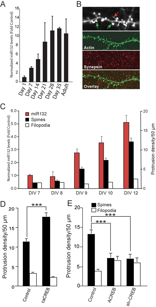
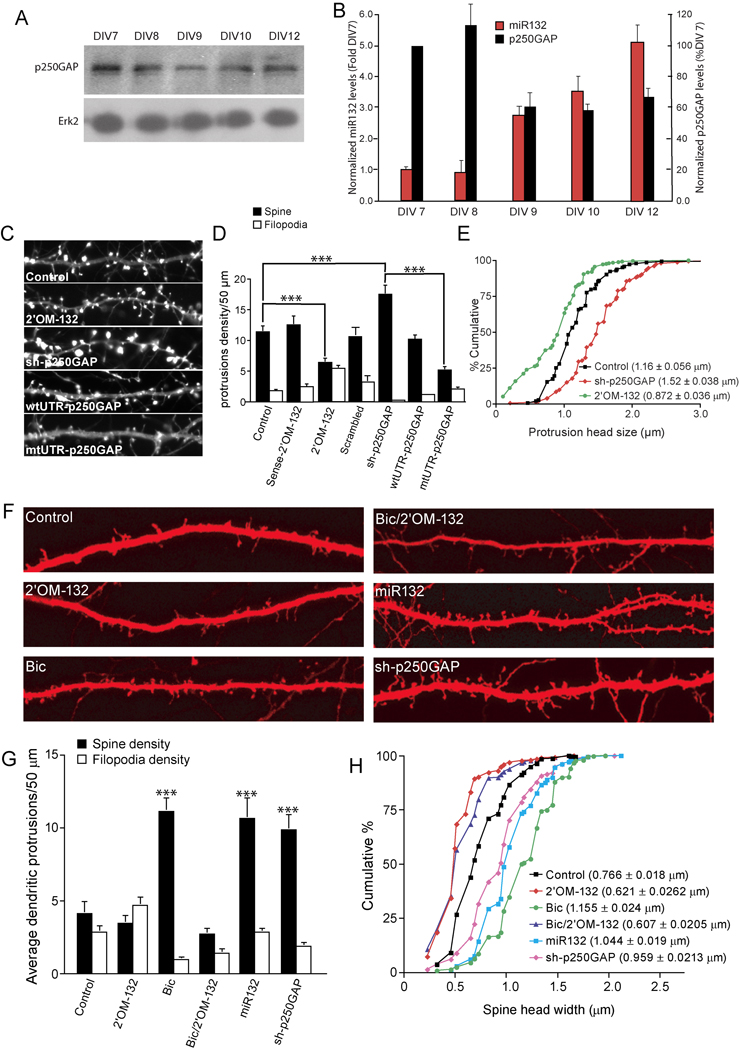
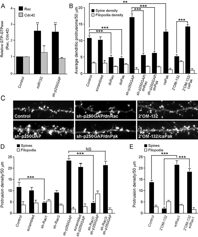
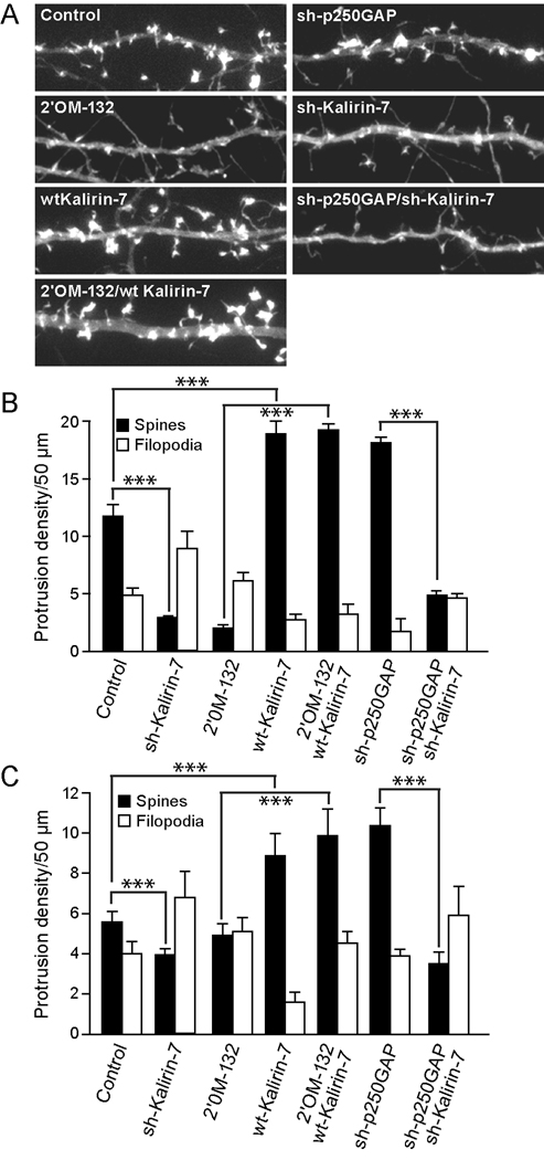
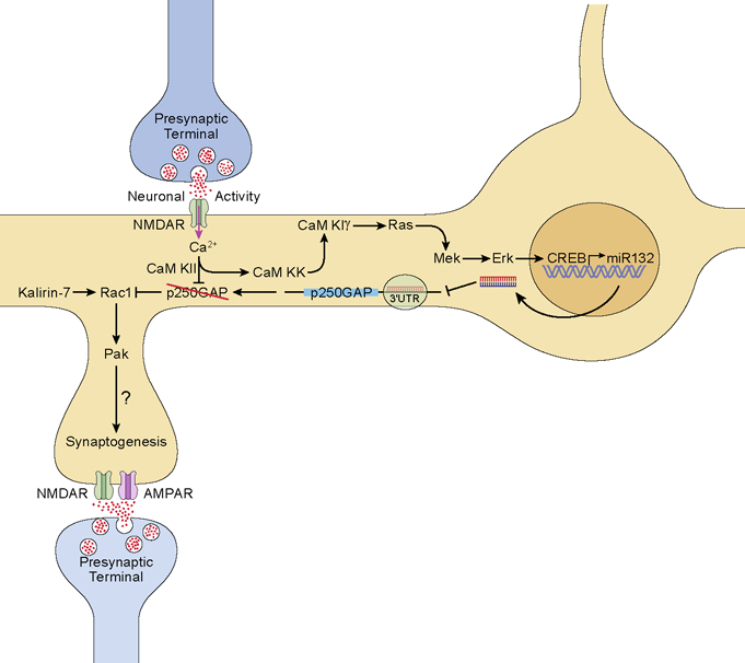
Activity-regulated gene expression is believed to play a key role in the development and refinement of neuronal circuitry. Nevertheless, the transcriptional networks that regulate synaptic plasticity remain largely uncharacterized. We show here that the CREB- and activity-regulated microRNA, miR132, is induced during periods of active synaptogenesis. Moreover, miR132 is necessary and sufficient for hippocampal spine formation. Expression of the miR132 target, p250GAP, is inversely correlated with miR132 levels and spinogenesis. Furthermore, knockdown of p250GAP increases spine formation while introduction of a p250GAP mutant unresponsive to miR132 attenuates this activity. Inhibition of miR132 decreases both mEPSC frequency and the number of GluR1-positive spines, while knockdown of p250GAP has the opposite effect. Additionally, we show that the miR132/p250GAP circuit regulates Rac1 activity and spine formation by modulating synapse-specific Kalirin7-Rac1 signaling. These data suggest that neuronal activity regulates spine formation, in part, by increasing miR132 transcription, which in turn activates a Rac1-Pak actin remodeling pathway.


Figures

Fig. 1
Fig. 1. miR132 expression correlates with spine formation in vivo and in vitro
(A) Mature miR132 levels are enhanced within the hippocampus during the period of afferent innervation. Hippocampi were isolated from rats (2 animals/developmental time point beginning at post natal day 1). RNA was isolated, reverse–transcribed, and analyzed by Taqman real-time PCR with miR132 cDNA primers. The data is normalized to GAPDH cDNA levels. (B) Cultured hippocampal neurons were transfected with YFP-γ actin to visualize dendritic protrusions, fixed on DIV 12, and immunostained for the presynaptic marker synapsin 1. (Upper panel) Spines (green arrows, protrusion head size 2 fold > shaft) were identified as mushroom-shaped projections with intense expression of EYFP-γ actin at their tips, whereas filopodia (red arrows, protrusion head size 2 fold ≤ shaft) were thin with low expression of EYFP-γ actin throughout. (C) Hippocampal neurons were transfected with EYFP-γ actin on DIV 6 and then cultured 7–12 days. At each time point, the cultures were either fixed and analyzed for spine and filopodia (black and white bars), or RNA was isolated and reverse transcribed and analyzed by Taqman-real time-PCR with mature-miR132 cDNA primers (red bars). For RT-PCR experiments, the data were normalized to GAPDH cDNA levels (± SEM n=5–6). (D, E) Hippocampal neurons were transfected with EYFP-γ actin ± empty vector (Control) ± ACREB ± siCREB or ± caCREB on DIV 7 and then fixed on DIV12. Dendrites were imaged, and at least three different 50 µm sections per neuron (25–30 neurons per condition) were analyzed. Quantitation of dendritic spines and filopodia are shown (± SEM, *** P < 0.001).
Fig. 2
Fig. 2. miR132 regulates spine formation by repressing p250GAP
(A) Developmentally timed hippocampal cultures were analyzed by Western blot for expression of p250GAP. (B) p250GAP expression was normalized to Erk2 levels and plotted with normalized mature-miR132 levels. (C–E) Hippocampal neurons DIV 7 were transfected with EYFP-γ actin and either empty vector (Control), Sense-2’OM-132 (sense control), 2’OM-132 (Antisense 2’OM-132), sh-p250GAP wtUTR-p250GAP, or mtUTR-p250GAP. On DIV12, the neurons were fixed and imaged. D,E, Effects of indicated transfections on (D) protrusion density and (E) spine head width. F–H, Hippocampal slices were cultured for 3 days and subjected to biolistic transfection with TFP ± empty vector (Control) or other plasmids as indicated. Slices were allowed to recover for 1 day and then stimulated (where indicated) with 20 µM bicuculline for 2 days. On DIV 7, dendritic protrusions were analyzed for spines and filopodia. (F) Representative examples of apical CA1 dendrites from pyramidal neurons in hippocampal organotypic sliced cultures are shown. Summary of the effects of transfection on protrusion density (G) and spine head width (H) are shown (*** denotes significance between test and control conditions). Statistical analyses utilized ANOVA and Tukey’s post-test. (± SEM, *** P < 0.001).
Fig. 3
Fig. 3. miR132 regulates synaptic function and morphology
(A, B) Effect of 2’OM-132 and sh-p250GAP on mEPSCs. Cultured neurons (DIV 7) were transfected with EYFP-γ actin and either empty vector (Control), 2’OM-132 or sh-p250GAP and mEPSCs were recorded on DIV12. (A) Representative traces of mEPSCs recorded from control, 2’OM-132, and sh-p250GAP transfected neurons. (B) Frequencies, amplitudes, rise times, and decay times of mEPSCs normalized to control. Statistical analyses utilized ANOVA and Tukey’s post-test. (± SEM, * P < 0.05). (C) Hippocampal neurons DIV 7 were transfected with mRFP-β actin and either empty vector (Control), 2’OM-132 or sh-p250GAP. The neurons were fixed on DIV 12 and stained for GluR1 using an N-terminal anti-GluR1 antibody. (D) Co-localization of GluR1 and actin-stained spines. (E) Hippocampal neurons DIV 7 were transfected with mRFP-β actin and either empty vector (Control) 2’OM-132 or sh-p250GAP. Neurons were fixed on DIV 12 and stained for the presynaptic protein, synapsin I. F, Effects of 2’OM-132 and sh-p250GAP on dendritic spines that show associated synapsin I staining. (± SEM, ** P < 0.01 , *** P < 0.001).
Fig. 4
Fig. 4. miR132 and p250GAP regulate the Rac-PAK pathway
Hippocampal cultures were transfected with myc-Rac or myc-Cdc42 ± empty vector (Control), miR132 or ± sh-p250GAP on DIV 5. On DIV7, Rac and Cdc42 activity were assayed using Rac/Cdc42 pull down assays. (A) Quantitation of Rac and Cdc42 activity from three independent experiments. (B, C) Rac and Pak activity is required for miR132 stimulated dendritic growth. Hippocampal neurons were transfected DIV 7 with plasmids encoding mRFP-β actin ± scrambled shRNA, miR132, sh-p250GAP, 2’OM-132, dnRac, dnPak, or caPak. Dendritic spine density was quantified on DIV 12. (D) Rac1 (and not Rac3) is downstream of p250GAP. Hippocampal neurons were transfected DIV 7 with plasmids encoding mRFP-β actin ± sh-p250GAP and, where indicated, shRNA constructs targeting Rac1 or Rac3 or a scrambled control. Dendritic spine density was quantified on DIV 12. Quantitation of dendritic spine and filopodia density is shown (± SEM). (E) Activation of Rac1 is sufficient to stimulate spine formation in the absence of miR132. Hippocampal neurons were transfected DIV 7 with plasmids encoding mRFP- actin ± 2’OM-132 and, where indicated, wild-type Rac1. Dendritic spine and filopodia density on DIV 12 is shown (± SEM, ** P < 0.01 , *** P < 0.001).
Fig. 5
Fig. 5. miR132-regulated spine formation requires the RacGEF, Kalirin-7
(A, B) Hippocampal neurons DIV 7 were transfected with mRFP-β actin and either empty vector (Control) 2’OM-132, sh-p250GAP, wtKalirin-7, sh-Kalirin-7, or combinations where indicated. On DIV12, the neurons were fixed and imaged. Representative dendrites are shown in A and quantified in B. (C) Hippocampal slices were cultured for 3 days and subjected to biolistic transfection with TFP ± other plasmids as indicated. Slices were allowed to recover for 1 day and then stimulated (where indicated) with 20 µM bicuculline for 2 days. On DIV 6, dendritic protrusions were analyzed for spines and filopodia (± SEM, *** P < 0.001).
Fig. 6
Fig. 6. Regulation of dendritic spine development by miR132
Increased synaptic activity stimulates glutamate release which, in turn, activates NMDA receptors. Ca2+ influx through the NMDAR activates CaMKII, CaMKK and CaMKIγ. CaMKIγ activates the MEK/Erk pathway to stimulate CREB-dependent synthesis of miR132. miR132 suppresses translation of p250GAP and CaMKII directly phosphorylates and inhibits p250GAP thereby releasing inhibition of Rac1. In the presence of Kalirin-7, Rac1 is activated and subsequently activates Pak to drive local actin polymerization and spine formation.

References

    1. Armstrong D, Dunn JK, Antalffy B, Trivedi R. Selective dendritic alterations in the cortex of Rett syndrome. J Neuropathol Exp Neurol. 1995;54:195–201. - PubMed
    1. Arthur JS, Fong AL, Dwyer JM, Davare M, Reese E, Obrietan K, Impey S. Mitogen- and stress-activated protein kinase 1 mediates cAMP response element-binding protein phosphorylation and activation by neurotrophins. J Neurosci. 2004;24:4324–4332. - PMC - PubMed
    1. Ashraf SI, McLoon AL, Sclarsic SM, Kunes S. Synaptic protein synthesis associated with memory is regulated by the RISC pathway in Drosophila. Cell. 2006;124:191–205. - PubMed
    1. Bahr BA. Long-term hippocampal slices: a model system for investigating synaptic mechanisms and pathologic processes. J Neurosci Res. 1995;42:294–305. - PubMed
    1. Bartsch D, Casadio A, Karl KA, Serodio P, Kandel ER. CREB1 encodes a nuclear activator, a repressor, and a cytoplasmic modulator that form a regulatory unit critical for long-term facilitation. Cell. 1998;95:211–223. - PubMed

Publication types

MeSH terms