Review
. 2009 Nov;25(11):501-10.
doi: 10.1016/j.tig.2009.09.011. Epub 2009 Oct 21.

Primary microcephaly: do all roads lead to Rome?

Affiliations
Review

Primary microcephaly: do all roads lead to Rome?

Gemma K Thornton et al. Trends Genet. 2009 Nov.

Abstract

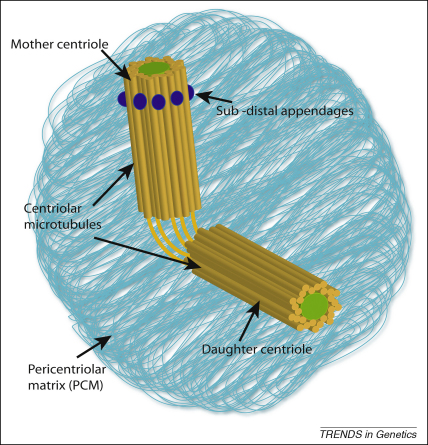
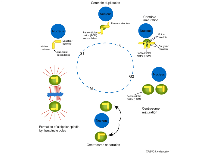
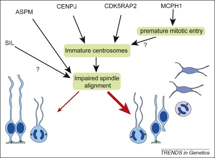
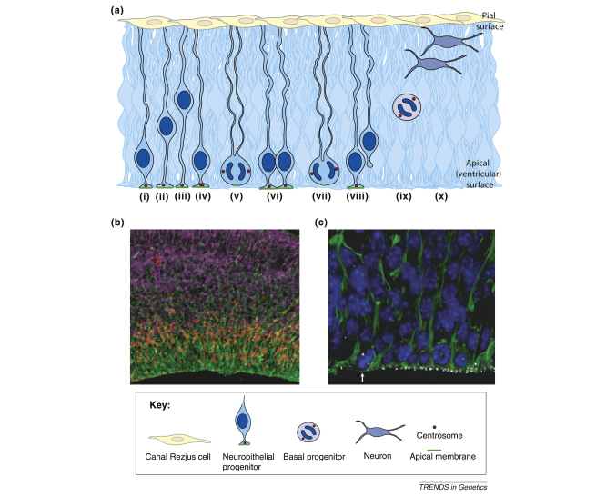
The relatively large brain and expanded cerebral cortex of humans is unusual in the animal kingdom and is thought to have promoted our adaptability and success as a species. One approach for investigating neurogenesis is the study of autosomal recessive primary microcephaly (MCPH), in which prenatal brain growth is significantly reduced without an effect on brain structure. To date, eight MCPH loci and five genes have been identified. Unexpectedly, all MCPH proteins are ubiquitous and localise to centrosomes for at least part of the cell cycle. Here, we focus on recent functional studies of MCPH proteins that reveal the centrosome as a final integration point for many regulatory pathways affecting prenatal neurogenesis in mammals.


Figures

Figure 1
Figure 1
The structure of the centrosome. The centrosome is a large complex structure. At its centre sit two centrioles, which are orientated perpendicularly to each other and are linked (orange lines). One centriole, termed the ‘mother’ centriole, is older and fully mature, whereas the other is called the ‘daughter’ centriole. The mother can be distinguished from the daughter by the presence of the sub-distal appendages (purple). Each centriole takes 1.5 cell cycles to reach maturation. The centrioles are barrel-like in appearance and are surrounded by a ninefold symmetrical arrangement of triplet microtubules (orange rods). The centriole pair accumulate numerous other proteins to form the PCM. The centrosome is one of the main microtubule organising centres in the cell, and is also implicated in coordinating many pathways.
Figure 2
Figure 2
The centrosome division cycle. Centrosome duplication and maturation are linked to cell-cycle progression. In early G1, post division, the cell contains one centriole pair consisting of a mature mother centriole, which has sub-distal appendages, and an immature daughter centriole, connected to each other by a linker. During G1, pro-centrioles form perpendicularly to both the mother and daughter centrioles and continue to lengthen as G1 progresses. Around S-phase, the original daughter centriole reaches maturation, acquiring sub-distal appendages, and the link between the original centriole pair is broken. PCM proteins begin to accumulate during centriole duplication, forming two centrosomes each containing a centriole pair. This accumulation continues through the G2 phase as centrosomes mature. The final steps in maturation are the addition of centriolar microtubules around late prophase. The centrosomes, which until now have remained associated with each other, separate and move to opposing sides of the nucleus. During mitosis a bipolar spindle forms to ensure faithful DNA segregation and, at each end, there is a spindle pole each containing a centrosome. The centrosome is responsible for generating the astral microtubule array, which enables correct spindle orientation to occur. Upon cytokinesis, each daughter cell inherits a single centriole pair and the cycle begins again.
Figure 3
Figure 3
A model suggesting how loss of the MCPH proteins could affect neurogenesis. Although the MCPH proteins act in diverse pathways, these can be shown to intersect, resulting in a common mechanism affecting neuron production. Loss of MCPH1 results in a shorter G1 phase of the cell cycle through premature mitotic entry, meaning that the centrosomes have not had sufficient time to mature before the onset of division. Deficiencies in CDK5RAP2 or CENPJ directly affect centrosome maturation (in the case of CENPJ loss, centrioles are no longer able to form). Immature centrosomes accumulate less PCM, and are also less able to generate astral microtubules. This is important because astral microtubules contact the cell cortex and provide information guiding spindle orientation during division. By contrast, ASPM and SIL localise specifically to mitotic spindle poles, where ASPM directly regulates spindle positioning. We speculate that SIL has a similar role. In apical NE progenitors, spindle positioning is tightly controlled to ensure bisection of the apical plasma membrane during symmetric division. By impairing spindle orientation even mildly, loss of any MCPH protein would lead to an increase in NE cells producing neurogenic progeny upon division (wide red arrow). The proportion of symmetric divisions is concomitantly reduced (thin red arrow), depleting the progenitor pool and limiting the total number of neurons that can be generated.
Figure I
Figure I
The developing mouse neuroepithelium. (a) The neuroepithelium and the process of cell division. Neuroepithelial cells (blue) have processes contacting the apical (ventricular) and pial (basal) surfaces. The nuclei in dark blue migrate basally during G1, cells (i,ii) undergo S phase at a basal position (iii); and migrate again apically during G2 (iv). The centrosomes (red circles) remain by the apical membrane (green). Mitosis occurs at the apical surface, where the centrosomes now form the spindle poles. Symmetrical division leads to the production of two identical neuroepithelial cells (v,vi). By contrast, asymmetrical division (vii) leads to production of one neuroepithelial cell, whereas the other daughter detaches from the membrane (viii) and becomes either a basal progenitor (ix) or a neuron. Basal progenitors (ix) lack processes and polarity and predominantly divide terminally to produce two neurons (x). (b) Immunofluorescence in E14 mouse neuroepithelium showing the apical progenitors and their processes (nestin, green), basal progenitors (Tbr2, red) and neurons (βIII Tubulin, purple). (c) Immunofluorescence in E12 mouse neuroepithelium showing the apical progenitors and their processes (nestin, green), the nuclei of the apical progenitors (DAPI, blue) and the centrosomes (γ-tubulin, white). Note the arrow pointing to the cell in metaphase.

References

    1. Cox J. What primary microcephaly can tell us about brain growth. Trends Mol. Med. 2006;12:358–366. - PubMed
    1. Nicholas A.K. The molecular landscape of ASPM mutations in primary microcephaly. J. Med. Genet. 2009;46:249–253. - PMC - PubMed
    1. Barkovich J.A. A developmental classification of malformations of the brainstem. Ann. Neurol. 1997;62:625–639. - PubMed
    1. Desir J. Primary microcephaly with ASPM mutation shows simplified cortical gyration with antero-posterior gradient pre- and post-natally. Am. J. Med. Genet. A. 2008;146A:1439–1443. - PubMed
    1. Saadi A. Compound heterozygous ASPM mutations associated with microcephaly and simplified cortical gyration in a consanguineous Algerian family. Eur. J. Med. Genet. 2008;52:180–184. - PubMed

MeSH terms

Substances