. 2010 Mar;51(3):1318-26.
doi: 10.1167/iovs.09-4455. Epub 2009 Oct 22.

Effects of human recombinant PEDF protein and PEDF-derived peptide 34-mer on choroidal neovascularization

Affiliations

Effects of human recombinant PEDF protein and PEDF-derived peptide 34-mer on choroidal neovascularization

Juan Amaral et al. Invest Ophthalmol Vis Sci. 2010 Mar.

Abstract

Purpose: Pigment epithelium-derived factor (PEDF) is a serpin with antiangiogenic properties. Previously, the authors showed that PEDF injected into the subconjunctiva reaches the choroid. Here, they examined the effects of PEDF polypeptide fragments on vessel sprouting and on choroidal neovascularization (CNV) after subconjunctival administration.

Methods: Recombinant human PEDF (rhuPEDF) was cleaved at its serpin-exposed loop by limited chymotrypsin proteolysis. Synthetic PEDF peptides 34-mer (Asp(44)-Asn(77)) and 44-mer (Val(78)-Thr(121)) were used. Ex vivo chick aortic vessel sprouting assays were performed. CNV was induced in rats by laser injury of Bruch's membrane. Daily subconjunctival injections (0.01-10 pmol/d protein) were performed for 5 days starting at day of injury or at the seventh day after injury. New vessel volumes were quantified using optical sections of choroid/RPE flat-mounts labeled with isolectin-Ib4. PEDF distribution was evaluated by immunofluorescence of choroid/RPE/retina cross-sections.

Results: Full-length rhuPEDF, cleaved rhuPEDF, or peptide 34-mer exhibited ex vivo antiangiogenic activity, but peptide 44-mer was inefficient. PEDF immunostaining around CNV lesions diminished after laser injury. Subconjunctival administration of rhuPEDF or 34-mer at 0.1 pmol/d decreased CNV lesion volumes by 52% and 47%, respectively, whereas those of 44-mer were similar to vehicle injections. Doses of 0.1 and 1 pmol/d rhuPEDF decreased fully developed CNV complex volumes by 45% and 50%, respectively, compared with vehicle injections.

Conclusions: A functional region for the inhibition of vessel sprouting and CNV resides within the 34-mer region of PEDF. Furthermore, subconjunctival administration of optimal range dosages of rhuPEDF or 34-mer can suppress and regress rat CNV lesions, demonstrating that these agents reach the choroid/RPE complex as functionally active molecules.


Figures

Figure 1.
Figure 1.
Inhibition vessel sprouting by PEDF. (A) Representative photographs of vessel sprouting in the presence and absence of 30 nM PEDF. ECGS (400 μg/mL) was used as an angiogenesis promoter. (B) Concentration response of the inhibitory activity of PEDF on vessel sprouting. Relative vessel sprouting was calculated from surface values of vessels and ring. Each point corresponds to the average of four replicate assays. The bars represent standard deviations. Closed circles: PEDF; closed squares: angiostatin (angiogenic inhibitor control). A plot of the relative vessel sprouting, represented by the vessel/ring surface ratio, as function of protein concentration is shown. Inset: concentration response of PEDF and angiostatin at lower concentrations (0–0.5 nM). Horizontal dotted line: negative control was without ECGS. Positive control was with ECGS but without PEDF or angiostatin.
Figure 2.
Figure 2.
Antiangiogenic activity of PEDF cleaved at its serpin-exposed loop. (A) Limited proteolysis of PEDF with chymotrypsin (CT +) at a protease/substrate ratio of 1:100 (wt/wt) incubated for 30 minutes at 25°C. Reactions were stopped with 0.5 mM AEBSF, a serine protease inhibitor, and products were resolved by SDS-PAGE. Coomassie blue–-stained 10% to 20% polyacrylamide gel is shown. M, molecular weight standards; PpB, phosphorylase B; BSA, bovine serum albumin; ova, ovalbumin; CA, carbonic anhydrase; Lys, lysozyme. (B) Effects of chymotrypsin-treated PEDF on vessel sprouting. In the x-axis, controls (negative was without ECGS; positive was with ECGS and without effectors) and effectors were additions of reaction mixtures after incubation at 25°C containing the concentrations of each indicated component (PEDF, chymotrypsin, and/or AEBSF) to rings with ECGS. The y-axis shows the relative area of vessel sprouting per ring (vessel/ring surface ratio). Each bar corresponds to the average of four replicates ± SD.
Figure 3.
Figure 3.
Effects of PEDF-derived peptides on vessel sprouting. (A) Representative photographs of vessel sprouting in the presence and absence of 10 nM peptide 34-mer. (B) Vessel sprouting assays with 1-, 10-, and 100-nM concentrations of PEDF-derived peptides (x-axis). Negative control was without ECGS; positive control was with ECGS and without peptides. Additions to rings with ECGS are indicated in the x-axis. The relative area of vessel sprouting (y-axis) as a function of peptide concentration was plotted. Each bar corresponds to the average of four replicate assays ± SD.
Figure 4.
Figure 4.
Immunolocalization of PEDF in laser-induced CNV. Retina/RPE/choroid cryosections from rats without laser injury and 1, 3, 5, and 7 days after injury stained with anti-PEDF primary antibodies followed by highly absorbed-Alexa 488 (PEDF, green) secondary antibodies. (blue, DAPI). ONL, outer nuclear layer.
Figure 5.
Figure 5.
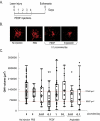
Full-length PEDF inhibited the growth of CNV complex lesions. (A) Scheme to illustrate the protocol for PEDF injections for the suppression of neovessel growth. (B) Representative flat-mount projections from confocal microscope Z-series 7 days after laser. The red channel identifies vessels (isolectin IB-4). Conditions are indicated below each projection. (C) Box-and-whisker plot representations (four experiments) of volume of CNV lesions from rats treated with daily administrations of effectors, as indicated on the x-axis. The y-axis represents neovessel lesion volume expressed in cubic micrometers. Each point corresponds to one CNV lesion (n = 163). Values inside the boxes correspond to the central 50% of measurements, their internal horizontal bars correspond to median values, and the vertical lines outside the boxes correspond to variances of measurements. The horizontal dotted red lines correspond to the median values (indicated to the right) of PBS and PEDF 0.1 pmol. *Significant or **highly significant difference compared with no injections and PBS.
Figure 6.
Figure 6.
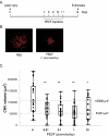
Full-length PEDF regressed preformed CNV complex lesions. (A) Scheme to illustrate protocol of PEDF injections for regression of fully formed neovesssels. (B) Representative flat-mount projections from epifluorescence microscope 14 days after laser. The red channel identifies vessels (isolectin IB-4). Conditions are indicated below each projection. (C) Box-and-whisker plot representations of volume of CNV lesions from rats treated with daily administrations of effectors, as indicated on the x-axis. The y-axis represents neovessel complex volume expressed in cubic micrometers. Each point corresponds to one CNV lesion (n = 143). Values inside the boxes correspond to the central 50% of measurements, their internal horizontal bars correspond to median values, and the vertical lines outside the boxes correspond to variances of measurements. The horizontal dotted red lines correspond to the median values (indicated to the right) of PBS and PEDF 1 pmol. *Significant or **highly significant difference compared with no injections and PBS.
Figure 7.
Figure 7.
Peptide 34-mer suppressed the growth of CNV complex lesions. (A) Representative flat-mount projections from confocal microscope 7 days after laser. The red channel identifies vessels (isolectin IB-4). Conditions are indicated below each projection. (B) Box-and-whisker plot representations of volume of CNV lesions from rats treated with daily administrations of effectors, as indicated on the x-axis. The y-axis represents neovessel complex volume expressed in cubic micrometers. Each point corresponds to one CNV lesion (n = 170). Values inside the boxes correspond to the central 50% of measurements, their internal horizontal bars correspond to median values, and the vertical lines outside the boxes correspond to variances of measurements. The horizontal dotted red lines correspond to the median values (indicated to the right) of PBS and PEDF 0.1 pmol. *Highly significant difference compared with PBS injections.

References

    1. Becerra SP. Focus on molecules: pigment epithelium-derived factor (PEDF). Exp Eye Res 2006;82:739–740 - PubMed
    1. Filleur S, Nelius T, de Riese W, Kennedy RC. Characterization of PEDF: a multi-functional serpin family protein. J Cell Biochem 2009;106:769–775 - PubMed
    1. Amaral J, Becerra S. Pigment Epithelium-Derived Factor and Angiogenesis: Therapeutic Implications Dordrecht, The Netherlands: Springer; 2008;311–337
    1. Bouck N. PEDF: anti-angiogenic guardian of ocular function. Trends Mol Med 2002;8:330–334 - PubMed
    1. Becerra SP. Structure-function studies on PEDF: a noninhibitory serpin with neurotrophic activity. Adv Exp Med Biol 1997;425:223–237 - PubMed

Publication types

MeSH terms