. 2009 Nov 3;106(44):18722-7.
doi: 10.1073/pnas.0908853106. Epub 2009 Oct 22.

Homozygous loss of BHD causes early embryonic lethality and kidney tumor development with activation of mTORC1 and mTORC2

Affiliations

Homozygous loss of BHD causes early embryonic lethality and kidney tumor development with activation of mTORC1 and mTORC2

Yukiko Hasumi et al. Proc Natl Acad Sci U S A. .

Abstract

Germline mutations in the BHD/FLCN tumor suppressor gene predispose patients to develop renal tumors in the hamartoma syndrome, Birt-Hogg-Dubé (BHD). BHD encodes folliculin, a protein with unknown function that may interact with the energy- and nutrient-sensing AMPK-mTOR signaling pathways. To clarify BHD function in the mouse, we generated a BHD knockout mouse model. BHD homozygous null (BHD(d/d)) mice displayed early embryonic lethality at E5.5-E6.5, showing defects in the visceral endoderm. BHD heterozygous knockout (BHDd(/+)) mice appeared normal at birth but developed kidney cysts and solid tumors as they aged (median kidney-lesion-free survival = 23 months, median tumor-free survival = 25 months). As observed in human BHD kidney tumors, three different histologic types of kidney tumors developed in BHD(d/+) mice including oncocytic hybrid, oncocytoma, and clear cell with concomitant loss of heterozygosity (LOH), supporting a tumor suppressor function for BHD in the mouse. The PI3K-AKT pathway was activated in both human BHD renal tumors and kidney tumors in BHD(d/+) mice. Interestingly, total AKT protein was elevated in kidney tumors compared to normal kidney tissue, but without increased levels of AKT mRNA, suggesting that AKT may be regulated by folliculin through post translational or post-transcriptional modification. Finally, BHD inactivation led to both mTORC1 and mTORC2 activation in kidney tumors from BHD(d/+) mice and human BHD patients. These data support a role for PI3K-AKT pathway activation in kidney tumor formation caused by loss of BHD and suggest that inhibitors of both mTORC1 and mTORC2 may be effective as potential therapeutic agents for BHD-associated kidney cancer.


Conflict of interest statement

The authors declare no conflict of interest.

Figures

Fig. 1.
Fig. 1.
Characterization of BHDd/d mouse embryo phenotype. (A) Embryos were isolated from BHD d/+ intercrosses, observed under a dissection microscope and genotyped by PCR. Numbers in parentheses represent embryos with abnormal appearance, as shown in (B and D). Gross appearance of BHDd/d (B and D) or BHD d/+ (C and E) embryos. H&E images of BHD +/+ (F), BHDd/+ (G), and BHDd/d (H and I) embryos at E6.5. Immunohistochemical staining of DAB2 was performed to see visceral endoderm of BHDd/+ (J), and BHDd/d (K) embryos. Magnified images of visceral endoderm of BHD+/+ (F' and L) and BHDd/d (H' and M) with H&E staining and DAB2 staining, respectively. Magnification; ×20 (F–I), ×40 (J and K), ×63 (F' and H'), and ×100 (L and M). [Scale bar, 100 μm (F–I) and 50 μm (J and K).]
Fig. 2.
Fig. 2.
Spontaneous kidney tumor development in BHDd/+ mice. BHDd/+ mice and BHD+/+ mice were aged and dissected randomly at different time points when they were moribund or had to be euthanized due to dermatitis. The renal capsule was removed from isolated kidneys and observed under dissection microscopy. (A) Gross appearance of BHDd/+ mouse kidney. (B) Solid tumor (**) in BHDd/+ kidney(*). (C) Kaplan-Meier analysis of BHDd/+ (n = 65) and BHD+/+ (n = 28) mice for kidney-lesion-free survival. Log-rank test (two-sided), P < 0.0001. Dotted lines, SEM. (D) Kaplan-Meier analysis of BHDd/+ (n = 65) and BHD+/+ (n = 28) mice for kidney-tumor-free survival (complex cysts and solid tumors). Log-rank test (two-sided), P = 0.0026. (E) The number of kidney lesions was counted in BHDd/+ mice between the ages of 20 and 25 months (n = 35). (Scale bar, SEM.)
Fig. 3.
Fig. 3.
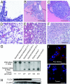
Histological analysis of BHDd/+ mouse kidney lesions. (A) H&E staining on formalin fixed paraffin embedded kidney samples. Cells lining the cyst show proliferative tubular epithelium unique to BHD cysts. (B) Cyst with papillary projection. (C) Micro solid tumors adjacent to BHD cyst. (D–F) Different histological features were observed in tumors, resembling clear cell (D), oncocytic hybrid (E), and oncocytoma (F). Magnification; ×5 (B), ×10 (C), and ×20 (A and D–F). [Scale bar, 400 μm (B); 200 μm (C); and 100 μm (A and D–F).] (G) Southern blotting was performed on genomic DNA isolated from normal kidneys and tumors. Loss of wild-type BHD alleles was observed in the mouse kidney tumors. (H) Western blotting was performed on the samples corresponding to (G), showing loss of FLCN protein expression. (I and J) Loss of FLCN protein in tumors was detected by immunostaining with the Duolink system. Representative staining of FLCN in adjacent kidney (I) and tumor (J) in the same BHDd/+ mouse. Magnification; ×100. (Scale bar, 20 μm.)
Fig. 4.
Fig. 4.
Elevated total AKT and phosphorylated AKT protein in kidney tumors arising in BHDd/+ mice. (A) Western blotting was performed on the protein lysates isolated from normal kidneys and kidney tumors. Both total AKT and phospho-AKT expressions were up-regulated in BHDd/+ tumors. Isoforms of AKT, AKT1, and AKT2, were overexpressed in tumors. (B) Immunohistochemical staining showed highly expressed p-AKT (Ser-473) in kidney tumors consistent with Western blot results. (C) qRT-PCR was performed on total RNA isolated from frozen tissue samples corresponding to (A). mRNA expression for AKT1 and AKT2 was not significantly different between tumors and normal kidneys. (D) Western blotting on tissue lysates corresponding to (A). AKT activation resulted in phosphorylation of downstream effectors of the AKT pathway in tumors. (E and F) Immunofluorescent staining of p-GSK3α/β was consistent with Western blotting in (D). Magnification; ×63. (Scale bar, 20 μm.)
Fig. 5.
Fig. 5.
mTOR pathway activation in kidney tumors from BHDd/+ mice. (A) Western blotting was performed on the protein lysates isolated from normal kidneys and kidney tumors. Both mTOR phosphorylation sites (Ser2448 and Ser2481) were more phosphorylated in tumors than in normal mouse kidneys. Downstream effectors of the mTOR pathway were highly expressed in mouse kidney tumors suggesting mTORC1 activity is up-regulated in these tumors. Immunofluorescence staining was performed on normal adjacent kidney, cysts, and tumors (B–G). Phospho-mTOR (Ser2448) (B–D) and its downstream target phospho-S6R (E–G) were more highly expressed in tumors and cysts lining cells than in adjacent kidney. Magnification, ×63. (Scale bar, 20 μm.)
Fig. 6.
Fig. 6.
Activation of the PI3K-AKT-mTOR signaling pathway in kidney tumors from human BHD patients. (A) Western blotting was performed on protein lysates of normal kidneys and kidney tumors from BHD patients, showing elevation of p-AKT, total AKT1, and total AKT2 in BHD tumors 1–3. (B–D) Protein expression levels of p-AKT(Ser-473) (B), p-S6K (Thr-421/Ser-424) (C), and p-S6R (Ser-240/244) (D) were quantified using the Meso Scale Discovery multiplex array system and were consistent with the Western blotting results. The protein expression levels are relative to the value of normal kidney 3, which is defined as 1. (E and F) Immunofluorescence staining of p-AKT (Ser-473) was performed on frozen sections of human BHD tumors (F) and adjacent normal kidneys (E). Magnification, ×40. (Scale bar, 50 μm.) (G) Information of BHD patients whose tumors were analyzed in this study.

References

    1. Zbar B, et al. Risk of renal and colonic neoplasms and spontaneous pneumothorax in the Birt-Hogg-Dubé syndrome. Cancer Epidemiol Biomarkers Prev. 2002;11:393–400. - PubMed
    1. Toro JR, et al. Birt-Hogg-Dubé syndrome: A novel marker of kidney neoplasia. Arch Dermatol. 1999;135:1195–1202. - PubMed
    1. Birt AR, Hogg GR, Dubé WJ. Hereditary multiple fibrofolliculomas with trichodiscomas and acrochordons. Arch Dermatol. 1977;113:1674–1677. - PubMed
    1. Nickerson ML, et al. Mutations in a novel gene lead to kidney tumors, lung wall defects, and benign tumors of the hair follicle in patients with the Birt-Hogg-Dubé syndrome. Cancer Cell. 2002;2:157–164. - PubMed
    1. Pavlovich CP, et al. Renal tumors in the Birt-Hogg-Dubé syndrome. Am J Surg Pathol. 2002;26:1542–1552. - PubMed

Publication types

MeSH terms