. 2010 Jan 1;285(1):713-22.
doi: 10.1074/jbc.M109.061838. Epub 2009 Oct 22.

Designed auto-assembly of nanostreptabodies for rapid tissue-specific targeting in vivo

Affiliations

Designed auto-assembly of nanostreptabodies for rapid tissue-specific targeting in vivo

Philippe Valadon et al. J Biol Chem. .

Abstract

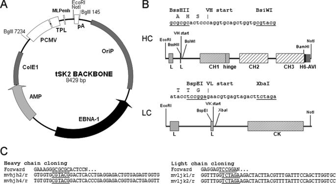
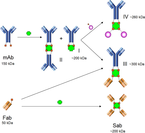
Molecular medicine can benefit greatly from antibodies that deliver therapeutic and imaging agents to select organs and diseased tissues. Yet the development of complex and defined composite nanostructures remains a challenge that requires both designed stoichiometric assembly and superior in vivo testing ability. Here, we generate nanostructures called nanostreptabodies by controlled sequential assembly of biotin-engineered antibody fragments on a streptavidin scaffold with a defined capacity for additional biotinylated payloads such as other antibodies to create bispecific antibodies as well as organic and non-organic moieties. When injected intravenously, these novel and stable nanostructures exhibit exquisite targeting with tissue-specific imaging and delivery, including rapid transendothelial transport that enhances tissue penetration. This "tinkertoy construction" strategy provides a very flexible and efficient way to link targeting vectors with reporter and/or effector agents, thereby providing virtually endless combinations potentially useful for multipurpose molecular and functional imaging in vivo as well as therapies.


Figures

FIGURE 1.
FIGURE 1.
tSK2 antibody expression vector series. A, backbone of the tSK2 episomal vector. Expression cassettes are inserted between the EcoRI site and the NotI site. PCMV, CMV promoter; TPL, adenovirus 5 tripartite leader sequence; MLPenh, adenovirus major late promoter enhancer; pA, polyadenylation site. B, heavy chain and light chain expression cassettes. Sequence of the cloning sites and translation before the start of variable regions are indicated above each diagram. L, leader peptide containing its own intron; H6, hexahistidine-tag; AVI, AviTag sequence. C, primer design. In the forward primers, N … indicates the start of the variable region sequence; sequence of the reverse primers is given for chains terminating by murine JH2, JH4, JK1, or JK2.
FIGURE 2.
FIGURE 2.
Antibody biotinylation. A, SEC-HPLC elugram of IMAC purified full-length bisbiotinylated chimeric antibody 833cb. B, Western analysis by SA-HRP conjugate of 833cb versus polyclonal human IgG; only the heavy chain is biotinylated as seen in reducing (R) conditions (NR, non-reducing conditions). C, expression of BirA in 293 cells (in green, left panel) follows a perinuclear membranous network that extends deep in the cytoplasm and substantially colocalizes with the Golgi network (in red, all panels); most of antibody-overexpressing 293 cells have an enlarged Golgi network staining that colocalizes with expression of 833cb (in green, right panel).
FIGURE 3.
FIGURE 3.
Influence of BirA concentration. The amount of BirA-expressing plasmid tSK2-BF was tested between 0 and 25% of the total plasmid content in the transfection mixture: A, concentration of 833cb antibody in the culture supernatant (ELISA), B, degree of antibody biotinylation by Western analysis using an SA-HRP conjugate (upper panel) and a human IgG1 reagent (bottom panel), and C, expression of BirA in 293 cells using an HA-antibody (upper panel) and an actin antibody (bottom panel).
FIGURE 4.
FIGURE 4.
Fab expression and streptabody formation. A, SEC-HPLC elugram of 833cb streptabody (Sab, red trace) prepared by incubating SA with a 5x excess of monobiotinylated 833cb Fab; remaining unbound Fab comigrates with the Fab alone (blue trace). B, titration of SA by increasing amounts of 833cb Fab; Fab/SA indicates the Fab to SA molar ratio and I, II, and III complexes between SA and one, two, and three Fabs, respectively.
FIGURE 5.
FIGURE 5.
Reactivity of bisbiotinylated antibodies. A, SEC-HPLC elugram of the reaction mixture of P27cb and SA at a 1:1 molar ratio. B, SEC-HPLC elugram of the reaction mixture of 833cb-link and SA at 1:1 molar ratio; Alexa Fluor 488 biocytin was added at the end of the reaction and fluorescence measurement on a 100-μl fraction collection is shown superimposed in arbitrary units. C, SEC-HPLC elugram of the reaction mixture of P27cb compound I with (red) or without (blue) 833cb Fab at a 1:2 molar ratio. D, Coomassie staining after SDS-PAGE in non-reducing conditions of: P27cb (lane 1), purified P27cb compound I (lane 2), purified P27cb compound III (lane 3), 833cb Fab (lane 4), and SA (lane 5). E, ELISA with GST-CAV1 (gray) or GST alone (white) coating and rat APP-HRP conjugate with the following primaries: nothing, P27cb Fab, 833cb Fab, P27cb, 833cb, monoSA/P27cb, monoSA/833cb, monoSA/P27cb-833cb Fab, monoSA/833cb-P27cb Fab, respectively from 1 to 9. F, same as E by dot-blots.
FIGURE 6.
FIGURE 6.
Targeting of 833 nanostreptabodies. A and B, in vivo CT-SPECT imaging of rats injected with 30 μCi of 833cb Sab obtained by reacting 125I-SA with 833cb Fab at a 4:1 molar ratio 10 min before injection (top panel) or 125I-SA alone (bottom panel). C, targeting of the 833cb Sab prepared with an Alexa Fluor 488 SA conjugate in rat lung tissue grown in a dorsal skinfold chamber in nude mice following tail vein injection (30 μg). D, time-space analysis of signal intensity on a vessel cross-section illustrating the transendothelial transport of 833cb Sab (37). E and F, lung targeting of the monoSa/833cb/mCherry complex analyzed by confocal microscopy in a rat lung, 250 μm, live section 1 h following tail vein injection of 400 μg (E) of the complex or 500 μg (F) mCherry alone; a few nonspecific large dots are visible on both images (yellow arrows) (bar, 50 μm, Z-stack, 10 μm).
FIGURE 7.
FIGURE 7.
Schematic representation of streptavidin/antibody complex formation. Bisbiotinylated mAbs react with SA (or avidin) to give the monoSA/antibody complex I and the monoSA/bisantibody complex II; monobiotinylated Fabs react with SA to give streptabodies (Sab) or with monoSA/antibodies I to give to bispecific antibodies III. MonoSA/antibody complex I reacts with biotinylated mCherry to give the red tripartite complex IV.

References

    1. Coloma M. J., Morrison S. L. (1997) Nat. Biotechnol. 15, 159–163 - PubMed
    1. Hust M., Jostock T., Menzel C., Voedisch B., Mohr A., Brenneis M., Kirsch M. I., Meier D., Dübel S. (2007) BMC Biotechnol. 7, 14. - PMC - PubMed
    1. Lee H. S., Shu L., De Pascalis R., Giuliano M., Zhu M., Padlan E. A., Hand P. H., Schlom J., Hong H. J., Kashmiri S. V. (1999) Mol. Immunol. 36, 61–71 - PubMed
    1. Marvin J. S., Zhu Z. (2005) Acta. Pharmacol. Sin. 26, 649–658 - PubMed
    1. Plückthun A., Pack P. (1997) Immunotechnology 3, 83–105 - PubMed

Publication types

LinkOut - more resources