

Neural prostheses and brain plasticity
- PMID: 19850976
- PMCID: PMC2935525
- DOI: 10.1088/1741-2560/6/6/065008
Neural prostheses and brain plasticity
Abstract
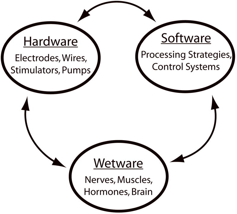
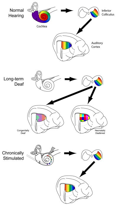
The success of modern neural prostheses is dependent on a complex interplay between the devices' hardware and software and the dynamic environment in which the devices operate: the patient's body or 'wetware'. Over 120 000 severe/profoundly deaf individuals presently receive information enabling auditory awareness and speech perception from cochlear implants. The cochlear implant therefore provides a useful case study for a review of the complex interactions between hardware, software and wetware, and of the important role of the dynamic nature of wetware. In the case of neural prostheses, the most critical component of that wetware is the central nervous system. This paper will examine the evidence of changes in the central auditory system that contribute to changes in performance with a cochlear implant, and discuss how these changes relate to electrophysiological and functional imaging studies in humans. The relationship between the human data and evidence from animals of the remarkable capacity for plastic change of the central auditory system, even into adulthood, will then be examined. Finally, we will discuss the role of brain plasticity in neural prostheses in general.
Figures
References
-
- Bao S, Chang EF, Woods J, Merzenich MM. Temporal plasticity in the primary auditory cortex induced by operant perceptual learning. Nat Neurosci. 2004;7:974–981. - PubMed
-
- Berthezene Y, Truy E, Morgon A, Giard MH, Hermier M, Franconi JM, Froment JC. Auditory cortex activation in deaf subjects during cochlear electrical stimulation. Evaluation by functional magnetic resonance imaging. Invest Radiol. 1997;32:297–301. - PubMed
-
- Blamey P, Arndt P, Bergeron F, Bredberg G, Brimacombe J, Facer G, Larky J, Lindstrom B, Nedzelski J, Peterson A, Shipp D, Staller S, Whitford L. Factors affecting auditory performance of postlinguistically deaf adults using cochlear implants. Audiology and Neuro-Otology. 1996;1:293–306. - PubMed
-
- Blamey PJ, Sarant JZ, Paatsch LE, Barry JG, Bow CP, Wales RJ, Wright M, Psarros C, Rattigan K, Tooher R. Relationships among speech perception, production, language, hearing loss, and age in children with impaired hearing. J Speech Lang Hear Res. 2001;44:264–285. - PubMed
-
- Busby PA, Clark GM. Gap detection by early-deafened cochlear-implant subjects. J Acoust Soc Am. 1999;105:1841–1852. - PubMed
Publication types
MeSH terms
Grants and funding
LinkOut - more resources
Full Text Sources
Medical