

doi: 10.1107/S1744309109036665.
Epub 2009 Sep 25.
High-resolution structure of human carbonic anhydrase II complexed with acetazolamide reveals insights into inhibitor drug design
Affiliations
- PMID: 19851004
- PMCID: PMC2765883
- DOI: 10.1107/S1744309109036665
Item in Clipboard
High-resolution structure of human carbonic anhydrase II complexed with acetazolamide reveals insights into inhibitor drug design
Acta Crystallogr Sect F Struct Biol Cryst Commun.
.
Abstract
The crystal structure of human carbonic anhydrase II (CA II) complexed with the inhibitor acetazolamide (AZM) has been determined at 1.1 A resolution and refined to an R(cryst) of 11.2% and an R(free) of 14.7%. As observed in previous CA II-inhibitor complexes, AZM binds directly to the zinc and makes several key interactions with active-site residues. The high-resolution data also showed a glycerol molecule adjacent to the AZM in the active site and two additional AZMs that are adventitiously bound on the surface of the enzyme. The co-binding of AZM and glycerol in the active site demonstrate that given an appropriate ring orientation and substituents, an isozyme-specific CA inhibitor may be developed.
Figures
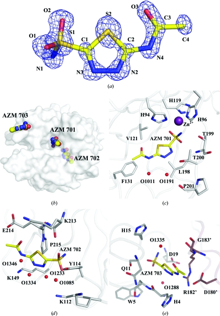
AZM. (a) The 2|F
o| − |F
c| electron-density map (blue) for the active-site AZM 701, contoured at 2.2σ. (b) Semi-transparent surface of CA II, showing the locations of the three AZMs. AZM–CA II interactions are shown in (c) for AZM 701 (active site), (d) for AZM 702 and (e) for AZM 703. Protein C atoms are coloured gray, symmetry-related protein C atoms mauve, ligand C atoms yellow, N atoms blue, O atoms red, S atoms orange and Zn atoms purple. Waters are represented by red spheres. This figure was created using PyMOL (DeLano, 2002 ▶).
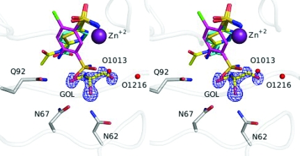
Stereo image of a superposition of inhibitors and glycerol for 3hs4 (ligand C atoms in yellow, protein C atoms in gray; this work), 2pov (C atoms in magenta; Alterio et al., 2007 ▶) and 2nno (C atoms in cyan; Srivastava et al., 2007 ▶). The 2|F
o| − |F
c| electron-density map (blue) of glycerol (GOL) is contoured at 2.0σ. N atoms are coloured blue, O atoms red, S atoms orange, Cl atoms green and Zn atoms purple. Waters are represented by red spheres. This figure was created using PyMOL (DeLano, 2002 ▶).
References
-
- Alterio, V., De Simone, G., Monti, S. M., Scozzafava, A. & Supuran, C. T. (2007). Bioorg. Med. Chem. Lett.17, 4201–4207. - PubMed
-
- Berman, H., Henrick, K. & Nakamura, H. (2003). Nature Struct. Biol.10, 980. - PubMed
-
- Breinin, G. M. & Gortz, H. (1954). AMA Arch. Ophthalmol.52, 333–348. - PubMed
-
- DeLano, W. L. (2002). The PyMOL Molecular Graphics System. http://www.pymol.org.
Publication types
MeSH terms
Substances
Grants and funding
LinkOut - more resources
Full Text Sources
Research Materials
