

Plasmacytoid dendritic cells: physiologic roles and pathologic states
- PMID: 19851130
- PMCID: PMC6329307
- DOI: 10.1097/PAP.0b013e3181bb6bc2
Plasmacytoid dendritic cells: physiologic roles and pathologic states
Abstract
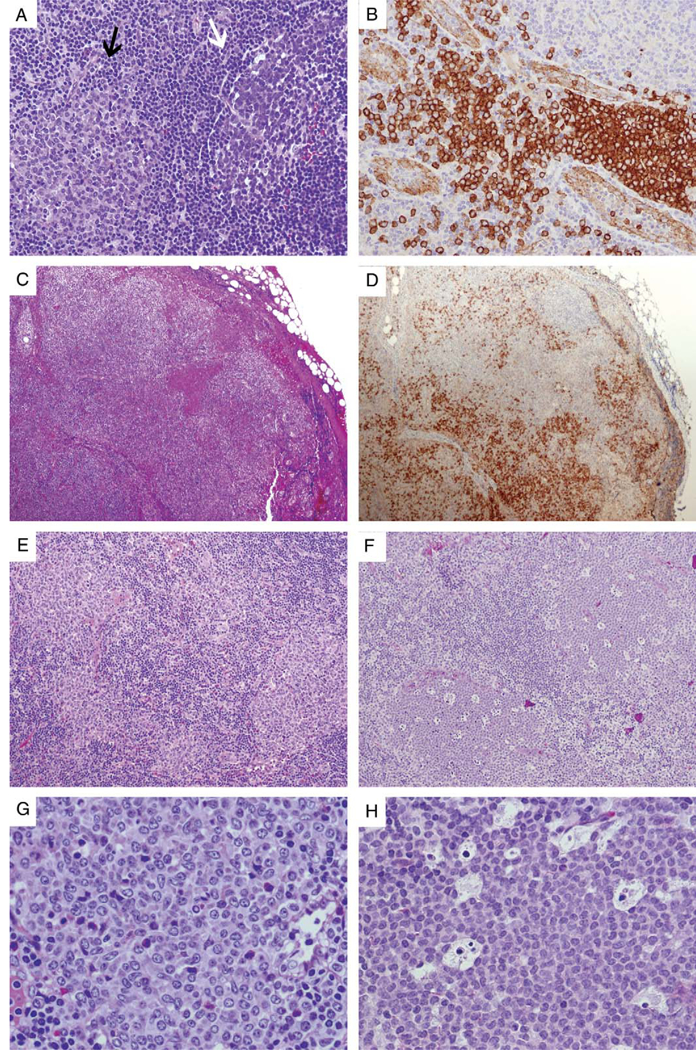
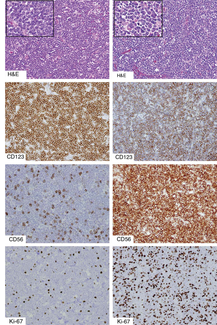
Plasmacytoid dendritic cells (PDCs) have perplexed pathologists for decades, undergoing multiple adjustments in nomenclature as their lineage and functions have been characterized. Although PDCs account for less than 0.1% of peripheral blood mononuclear cells, they serve as a principal source of interferon-alpha and are also known as interferon-I producing cells (IPCs). Upon activation in vitro, they can differentiate into dendritic cells, and recent studies have substantiated a potential role in antigen presentation. Thus, PDCs may act as a link between innate and adaptive immunity. Normally found in small quantities in primary and secondary lymphoid organs, PDCs accumulate in a variety of inflammatory conditions, including Kikuchi-Fujimoto lymphadenopathy, hyaline-vascular Castleman disease, and autoimmune diseases, and in certain malignancies such as classical Hodgkin lymphoma and carcinomas. Demonstrating potential for neoplastic transformation reflective of varying stages of maturation, clonal proliferations range from PDC nodules most commonly associated with chronic myelomonocytic leukemia to the rare but highly aggressive malignancy now known as blastic plasmacytoid dendritic cell neoplasm (BPDCN). Formerly called blastic natural killer cell lymphoma or CD4/CD56 hematodermic neoplasm, BPDCN, unlike natural killer cell lymphomas, is not associated with Epstein-Barr virus infection and is generally not curable with treatment regimens for non-Hodgkin lymphomas. In fact, this entity is no longer considered to be a lymphoma and instead represents a unique precursor hematopoietic neoplasm. Acute leukemia therapy regimens may lead to sustained clinical remission of BPDCN, with bone marrow transplantation in first complete remission potentially curative in adult patients.
Figures
References
-
- Lennert K, Remmele W. Karyometrische Untersuchungen an Lymphknotenzellen des Menschen: i mitt germinoblasten, lymphoblasten und lymphozyten. Acta Haematol (Basel) 1958;19:99–113. - PubMed
-
- Müller-Hermelink HK, Kaiserling E, Lennert K. Pseudofolli- kuläre Nester von Plasmazellen (eines besonderen Typs?) in der paracorticalen Pulpa menschlicher Lymphknoten. Virchows Arch (Cell Pathol). 1973;14:47–56. - PubMed
-
- Feller AC, Lennert K, Stein H, et al. Immunohistology and etiology of histiocytic necrotizing lymphadenitis. Report of three instructive cases. Histopathology. 1983;7:825–839. - PubMed
Publication types
MeSH terms
Substances
Grants and funding
LinkOut - more resources
Full Text Sources
Research Materials
