. 2010 Jan 6;29(1):145-57.
doi: 10.1038/emboj.2009.308. Epub 2009 Oct 22.

The helicases DinG, Rep and UvrD cooperate to promote replication across transcription units in vivo

Affiliations

The helicases DinG, Rep and UvrD cooperate to promote replication across transcription units in vivo

Hasna Boubakri et al. EMBO J. .

Erratum in

  • EMBO J. 2010 Jan 6;29(1):278

Abstract

How living cells deal with head-on collisions of the replication and transcription complexes has been debated for a long time. Even in the widely studied model bacteria Escherichia coli, the enzymes that take care of such collisions are still unknown. We report here that in vivo, the DinG, Rep and UvrD helicases are essential for efficient replication across highly transcribed regions. We show that when rRNA operons (rrn) are inverted to face replication, the viability of the dinG mutant is affected and over-expression of RNase H rescues the growth defect, showing that DinG acts in vivo to remove R-loops. In addition, DinG, Rep and UvrD exert a common function, which requires the presence of two of these three helicases. After replication blockage by an inverted rrn, Rep in conjunction with DinG or UvrD removes RNA polymerase, a task that is fulfilled in its absence by the SOS-induced DinG and UvrD helicases. Finally, Rep and UvrD also act at inverted sequences other than rrn, and promote replication through highly transcribed regions in wild-type E. coli.


Conflict of interest statement

The authors declare that they have no conflict of interest.

Figures

Figure 1
Figure 1
Schematic representation of the inverted region in the mutants InvA (top) and InvBE (bottom). Numbers indicate the sequence coordinates in the wild-type E. coli MG1655 chromosome. The large black arrows indicate the inversion end points (lambda att sites). The grey arrows indicate the position of rrn operons (the coordinates of rrnA, and of rrnE and rrnB 3′ ends are indicated). The vertical arrows show the position of NotI sites (used for PFGE).
Figure 2
Figure 2
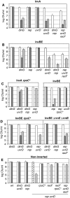
The helicases Rep, UvrD and DinG are required for colony formation in Inv mutants. Appropriate dilutions of overnight cultures at 37°C in MM (OD 1.0–1.5) were plated on MM and LB plates, which were incubated at 37°C. Unmarked positions on the left of (A) (InvA), (B) (InvBE) and (C) (InvABE) are data points for Inv mutants that express all helicases. White boxes: colony forming units (cfu)/ml on MM plates after 48 h incubation; dark grey boxes: cfu/ml on LB plates after 16–24 h incubation; light grey boxes: cfu/ml on LB plates after 48 h of incubation. The hatched box indicates cfu/ml on MM after 3 days incubation. The results are also presented in Supplementary Table S2.
Figure 3
Figure 3
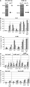
rep, uvrD and/or dinG mutations prevent Inv-fragment migration in PFGE. (A) InvA dinG rep cells (left panel) or InvBE rep cells (right panel) were propagated in MM or in LB for 1 or 2 h as indicated above each lane. Cells were lysed in plugs, chromosomes were treated with NotI, and restriction fragments were separated by PFGE. As the InvA-fragment is 50 kb and the InvBE-fragment is 138 kb (Figure 1), different migration conditions were used for InvA and InvBE mutants. For each panel, left lanes show the Et Br stained gel, the position of the wells and of the Inv fragment is indicated; right lanes Southern hybridization with a probe that detects the Inv-fragments after DNA transfer to a nylon membrane. (BE) Percentage of Inv-fragment DNA retained in wells for various mutant strains, quantified after Southern hybridization. Unmarked positions on the left of (A) (InvA) and (B) (InvBE) are data points for Inv mutants that express all helicases. White boxes: percentage of non-migrating Inv fragment in cells grown in MM; dark grey boxes: percentage of non-migrating Inv fragment in cells grown in LB for 1 h; light grey boxes: percentage of non-migrating Inv fragment in cells grown in LB for 2 h. The results are also presented in Supplementary Table S4.
Figure 4
Figure 4
Replication forks are arrested in inverted rrn. 2D gels were used to examine DNA replication in restriction fragments containing a large 3′ region of rrnA in InvA mutants and of rrnE in InvBE mutants. (A) Schematic representation of the restriction fragments used for 2D gels, left InvA, right InvBE. Top line, the position of rrn and of restriction sites are shown; bottom lines, schematic representation of the forked fragments when replication is arrested at the 5′ end of rrn, distances from the restriction sites to the 5′ end of rrn and the relative size of the forked fragments compared with linear fragments are indicated. (B, C) DNA from various InvA (B) and InvBE (C) mutants were digested with the indicated restriction enzyme, analysed by 2D gels and probed for the sequence just downstream of the analysed rrn. The left panel shows a simulation of replication arrest in the entire restriction fragment with an increased arrest in about 500 pb around the rrn transcription terminator sequence. The mutants used are indicated above each panel. In the InvBE rep and rep dinG rpoC* mutants, the signal of increased replication arrest in the transcription termination region was not always observed, independently of the restriction enzyme used, and one example of each situation is shown.
Figure 5
Figure 5
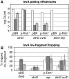
Inv dinG mutants are rescued by Rnase H overproduction, but not other mutants. (A) Plating efficiencies of InvA mutants [pEM-Ap] overnight cultures determined as in Figure 2. (B) Percentage of InvA-fragment retained in PFG wells in pEM-Ap containing cells determined as in Figure 3.
Figure 6
Figure 6
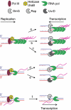
Rescue of transcription-blocked replication forks by helicases. Schematic representation of a replication fork blocked by a transcription unit. Top, a replication fork encounters an oppositely oriented highly expressed rrn operon (Inv mutant in LB): (a) in cells proficient for all helicases and in a uvrD single mutant, Rep translocating towards the transcription unit on the leading strand template and DinG on the lagging strand template act in concert; (b) in a rep mutant both UvrD and DinG are required, UvrD translocating on the leading strand template and DinG on the lagging strand template act in concert; (c) in a dinG mutant, both Rep and UvrD are required, they both translocate towards the transcription unit on the leading strand template; because R-loops form on the lagging strand template (not shown) where no helicase is present, R-loops are deleterious. Middle, a replication fork encounters an oppositely oriented moderately expressed rrn operon (Inv mutant in MM): (d) Rep only (dinG uvrD mutant) or (e) UvrD only (dinG rep mutant)is sufficient for replication. Bottom, a replication fork encounters a normally oriented (co-directional) moderately expressed rrn operon (wild-type chromosome in MM): this is the only condition in which DinG alone (rep uvrD recF mutant) allows full viability. Full lines: template DNA; dashed lines: newly synthesized DNA; oval: replisome; yellow circles: DnaB helicase; green indented circles: RNA Pol; pink lines: rRNA. Helicases are shown as grey indented circles: hatched DinG, light grey Rep, dark grey UvrD.

Similar articles

Cited by

References

    1. Appleman JA, Ross W, Salomon J, Gourse RL (1998) Activation of Escherichia coli rRNA transcription by FIS during a growth cycle. J Bacteriol 180: 1525–1532 - PMC - PubMed
    1. Azvolinsky A, Dunaway S, Torres JZ, Bessler JB, Zakian VA (2006) The S. cerevisiae Rrm3p DNA helicase moves with the replication fork and affects replication of all yeast chromosomes. Genes Dev 20: 3104–3116 - PMC - PubMed
    1. Bartlett MS, Gaal T, Ross W, Gourse RL (1998) RNA polymerase mutants that destabilize RNA polymerase-promoter complexes alter NTP-sensing by rrn P1 promoters. J Mol Biol 279: 331–345 - PubMed
    1. Bartlett MS, Gaal T, Ross W, Gourse RL (2000) Regulation of rRNA transcription is remarkably robust: FIS compensates for altered nucleoside triphosphate sensing by mutant RNA polymerases at Escherichia coli rrn P1 promoters. J Bacteriol 182: 1969–1977 - PMC - PubMed
    1. Bedinger P, Hochstrasser M, Jongeneel CV, Alberts BM (1983) Properties of the T4 bacteriophage DNA replication apparatus: the T4 dda DNA helicase is required to pass a bound RNA polymerase molecule. Cell 34: 115–123 - PubMed

Publication types

LinkOut - more resources