. 2009 Dec 2;28(23):3667-80.
doi: 10.1038/emboj.2009.302. Epub 2009 Oct 22.

Optimal function of the DNA repair enzyme TDP1 requires its phosphorylation by ATM and/or DNA-PK

Affiliations

Optimal function of the DNA repair enzyme TDP1 requires its phosphorylation by ATM and/or DNA-PK

Benu Brata Das et al. EMBO J. .

Abstract

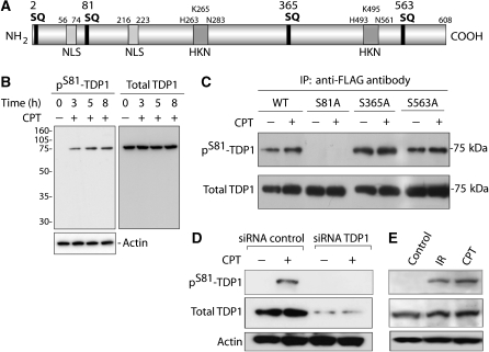
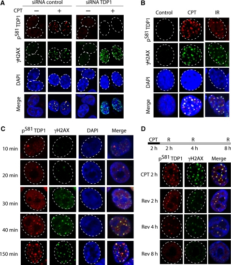
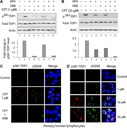
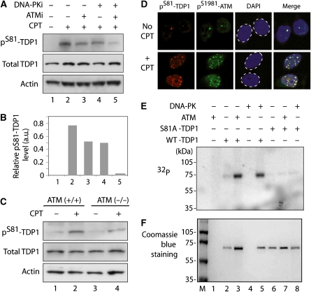
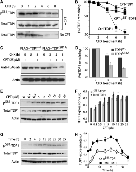
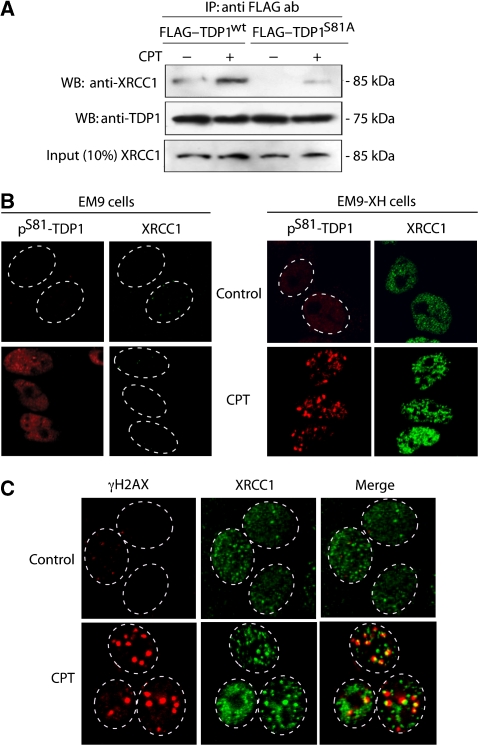
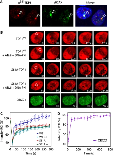
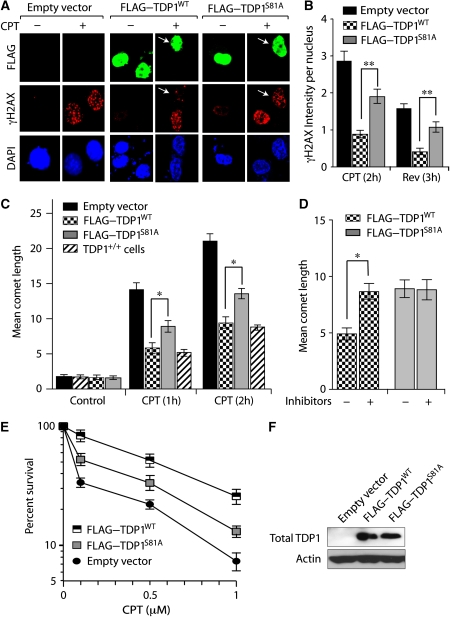
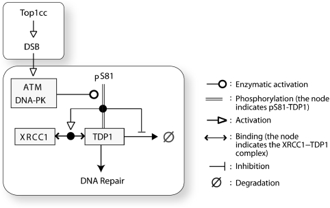
Human tyrosyl-DNA phosphodiesterase (TDP1) hydrolyzes the phosphodiester bond at a DNA 3' end linked to a tyrosyl moiety. This type of linkage is found at stalled topoisomerase I (Top1)-DNA covalent complexes, and TDP1 has been implicated in the repair of such complexes. Here we show that Top1-associated DNA double-stranded breaks (DSBs) induce the phosphorylation of TDP1 at S81. This phosphorylation is mediated by the protein kinases: ataxia-telangiectasia-mutated (ATM) and DNA-dependent protein kinase (DNA-PK). Phosphorylated TDP1 forms nuclear foci that co-localize with those of phosphorylated histone H2AX (gammaH2AX). Both Top1-induced replication- and transcription-mediated DNA damages induce TDP1 phosphorylation. Furthermore, we show that S81 phosphorylation stabilizes TDP1, induces the formation of XRCC1 (X-ray cross-complementing group 1)-TDP1 complexes and enhances the mobilization of TDP1 to DNA damage sites. Finally, we provide evidence that TDP1-S81 phosphorylation promotes cell survival and DNA repair in response to CPT-induced DSBs. Together; our findings provide a new mechanism for TDP1 post-translational regulation by ATM and DNA-PK.


Conflict of interest statement

The authors declare that they have no conflict of interest.

Figures

Figure 1
Figure 1
Human TDP1 is phosphorylated at S81 on DNA Damage. (A) Schematic representation of human TDP1 with the four SQ motifs. The positions of the active sites (HKN motifs) and nuclear localization sequences (NLS) are also shown. (B) In response to CPT treatment, the phospho-specific antibody recognizes a single band with molecular weight corresponding to TDP1. HCT116 cells were treated with 25 μM CPT for the indicated times and cellular extracts were analysed by western blotting. The blot shown on the left was stripped and re-probed with anti-TDP1 antibody (right). (C) Detection of pS81 by the phospho-specific antibody. Flag-tagged wild-type (WT) and mutant (S81A, S365A, and S563A) TDP1 were ectopically expressed in HCT116 cells. After treatment with 25 μM CPT for 2 h, FLAG–TDP1 was immunoprecipitated using anti-FLAG antibody and immune complexes were blotted with the anti-pS81-TDP1 antibody. The same blot was stripped and re-probed with anti-TDP1 antibody. The pS81-TDP1 signal in the untreated samples (−) is probably related to transfection. (D) SiRNA knockdown of TDP1 in MDA MB 231 cells abrogates the pS81-TDP1 signal on DNA damage with CPT (25 μM for 2 h), indicating the specificity of the antibody for pS81 of TDP1. (E) Phosphorylation of endogenous TDP1 at S81 in HCT116 cells after treatment with IR (10 Gy) and CPT (25 μM). Cells were analysed 3 h after irradiation or continuous CPT exposure.
Figure 2
Figure 2
TDP1 phosphorylation at S81 is associated with its focal accumulation at DNA damage sites. (A) Immunofluorescence microscopy analysis of TDP1 knockdown cells treated with CPT (25 μM for 2 h). Focal accumulation of pS81-TDP1 and γH2AX are shown in red and green, respectively. Nuclei were stained using DAPI. Note the co-localization of pS81-TDP1 and γH2AX (merged images). (B) Immunofluorescence microscopy analysis of HCT116 cells treated with CPT (25 μM for 3 h) or IR (10 Gy+3 h). Focal accumulations of pS81-TDP1 and γH2AX are shown in red and green, respectively. Nuclei were stained with DAPI. Co-localization of pS81-TDP1 and γH2AX (merged images) indicates the formation of pS81-TDP1 foci at sites of DNA damage. (C) Kinetics of pS81-TDP1 foci in HCT116 cells treated with CPT (25 μM). pS81-TDP1 and γH2AX foci are shown in red and green, respectively. Nuclei were stained using DAPI. (D) Kinetics of disappearance of pS81-TDP1 foci after CPT removal. HCT116 cells were treated with CPT (25 μM for 2 h). After CPT removal (R), cells were cultured in drug-free medium for the indicated times (indicated at the top). Nuclei are outlined in dashed white lines.
Figure 3
Figure 3
TDP1 phosphorylation at S81 is induced both by replication- and transcription-associated DNA damage. (A, B) Top panels: HCT116 cells were pre-treated with 1 μM aphidicolin (APH) for 15 min or 100 μM of DRB for 1 h followed by 2-h treatment with CPT at (A) low (1 μM) or (B) high concentration (25 μM) to prevent the replication- and transcription-associated DSB, respectively. Cells were examined for pS81-TDP1 and total TDP1 by western blotting. Bottom panels: densitometry analysis of pS81-TDP1 normalized against actin represents the relative level of pS81-TDP1. The numbers correspond to lanes in the top panels. (C) Immunofluorescence analysis of human lymphocytes treated with CPT (1 μM for 2 h). Nuclei were stained using DAPI. Lymphocytes were pre-treated with 100 μM of DRB for 1 h to arrest transcription, followed by a treatment with CPT for 2 h. Co-localization of pS81-TDP1 (red) and γH2AX (green) indicates that pS81-TDP1 foci are formed at damage sites. (D) Representative pictures showing dose-dependent increase of pS81-TDP1 staining with increasing concentrations of CPT for 2 h.
Figure 4
Figure 4
Both ATM and DNA-PK are involved in TDP1 phosphorylation at S81. (A) Effect of the ATM inhibitor KU55933 (ATMi) and of the DNA-PK inhibitor NU7441 (DNA-PKi) on TDP1 phosphorylation at S81. HCT116 cells were treated with inhibitors (10 μM for 1 h) before the addition of CPT (25 μM for 2 h). TDP1 phosphorylation (pS81-TDP1) and total TDP1 were analysed by western blotting. (B) Densitometry analysis of pS81-TDP1 normalized to actin under the indicated conditions. Numbers refer to the lanes in (A). (C) Defective phosphorylation of TDP1 at S81 in human A-T fibroblasts on treatment with CPT (25 μM for 2 h). The response of isogenic ATM-complemented A-T cells (+/+) is also shown. (D) Co-localization of pS81-TDP1 (red) and pS1981-ATM (green) foci in HCT116 cells treated with CPT (25 μM for 2 h). Nuclei stained with DAPI are outlined in dashed lines. (E) In vitro kinase assays with ATM or DNA-PK immunoprecipitated from HCT116 cells. The substrates were His-tagged TDP1: wild type (WT) and S81A. (F) Coomassie blue staining showing the amount of substrate in each reaction.
Figure 5
Figure 5
S81 stabilizes TDP1. (A, B) Increased half-life of endogenous TDP1 on CPT treatment. HCT116 cells were treated with 25 μM CPT for 2 h immediately followed by a treatment with cycloheximide (CHX) for the indicated times. Proteins (pS81-TDP1 and total TDP1) were (A) detected by western blotting (representative experiment) and (B) quantified by densitometry. (C, D) HCT116 cells were transfected with wild-type (WT) or the phospho-mutant S81A–FLAG-tagged TDP1, and 24 h later were treated with 25 μM CPT for 2 h immediately followed by a treatment with CHX. Protein levels were (C) determined by western blotting and (D) quantified by densitometry. (E) HCT116 cells were treated with increasing concentrations of CPT (0.1–25 μM) for 3 h and protein levels were analysed by western blotting (representative experiment is shown). (F) Densitometry analysis of pS81-TDP1 and total TDP1 normalized to actin represents the relative level of total and pS81-TDP1 after treatment with the indicated CPT concentrations. (G) Kinetics of TDP1 accumulation in HCT116 cells treated with 1 μM CPT for the indicated times. (H) Densitometry analysis of pS81-TDP1 and total TDP1 normalized to actin. Data represent the mean±s.e. values of independent experiments.
Figure 6
Figure 6
Phosphorylation of TDP1 at S81 promotes TDP1 binding to XRCC1. (A) Wild-type (WT) and the phospho-mutant (S81A), FLAG-tagged TDP1 were ectopically expressed in HCT116 cells. After CPT treatment (25 μM for 2 h), ectopic TDP1 was immunoprecipitated using anti-FLAG antibody and the immune complexes were blotted with anti-XRCC1 antibody. The same blot was stripped and re-probed with anti-TDP1 antibody. Aliquots (10%) of the input show the uniform presence of XRCC1 before immunoprecipitation. (B) XRCC1-deficient (EM9) and EM9 cells stably transfected with human XRCC1 (EM9-XH) were treated with CPT (5 μM for 2 h). Immunofluorescence microscopy shows significant reduction in pS81-TDP1 foci (red) in EM9 cells, whereas focal accumulation is restored in EM9-XH cells (green). (C) Immunofluorescence microscopy analysis of HCT116 cells treated with CPT (25 μM for 2 h). Focal accumulations of XRCC1 and γH2AX are shown in green and red, respectively. Nuclei are outlined in dashed white lines. Partial co-localization of XRCC1 and γH2AX (merged images) indicates subsets of CPT-induced XRCC1 foci at CPT-induced DSB sites.
Figure 7
Figure 7
Recruitment of RFP–TDP1 to DNA damage sites depends on S81. (A) Immunochemical detection of endogenous TDP1 phosphorylation at S81 after laser-induced DNA damage. Cells were fixed after 10 min of microirradiation and were stained with anti-pS81-TDP1 (left) or γH2AX antibody (middle). Nuclei were stained using DAPI. Co-localization of pS81-TDP1 and γH2AX (merged images) indicates the accumulation of pS81-TDP1 at sites of laser-induced DNA damage sites. Arrows indicate the sites of irradiation. (B) Recruitment of TDP1WT–RFP, phosphomutant TDP1S81A–RFP, XRCC1–YFP, TDP1WT–RFP, and TDP1S81A–RFP, pre-treated for 1 h with both the ATM inhibitor KU55933 (ATMi, 10 μM) and the DNA-PK inhibitor NU7441 (DNA-PKi, 10 μM), were analysed by microirradiation using live-cell microscopy and photobleaching (FRAP analysis). A sub-nuclear spot indicated by a circle was bleached (BLH) for 300 ms and photographed at regular intervals of 10 s thereafter. Successive images taken for ∼300 s after bleaching illustrate the level of return of fluorescence into the bleached areas. For XRCC1–YFP, images were photographed for ∼800 s at intervals of 60 s. (C) Quantitative FRAP data of HCT 116 cells expressing TDP1WT–RFP, TDP1WT–RFP (ATMi+DNA-PKi) or the phosphomutant TDP1S81A–RFP and TDP1S81A–RFP (ATMi and DNA-PKi) (n=3) showing mean curves. Error bars represent the s.e. values of the mean. (D) Quantitative FRAP data of HCT116 cells expressing XRCC1–YFP (n=5). Error bars represent the s.e.m. values.
Figure 8
Figure 8
S81-TDP1 promotes cell survival and DNA repair in response to CPT-induced DNA damage. (A) Immunofluorescence microscopy analysis for induction of γH2AX in TDP1−/− cells expressing FLAG–TDP1WT, FLAG–TDP1S81A or vector control treated with CPT (5 μM for 2 h). Representative pictures showing expression of FLAG–TDP1WT or FLAG-TDP1S81A detected by immunofluorescence staining with anti-FLAG antibody (green). γH2AX induction is shown in red. Cells were counterstained with DAPI to visualize nuclei. Arrows indicate the differential induction of γH2AX in TDP1−/− cells transfected with TDP1WT or TDP1S81A. The level of γH2AX is also shown in the untransfected (−TDP1WT, −TDP1S81A) cells in the same field 24 h after of transfection. (B) Quantification of γH2AX intensity per nucleus after CPT treatment (5 μM for 2 h) and 3 h after CPT removal (5 μM CPT for 2–3 h in drug-free medium) was calculated for 30–40 cells (calculated value±s.e.). Asterisks denote significant difference (**P<0.001; t test). (C) Differential DNA strand breaks measured by alkaline comet assays in response to CPT (5 μM) in TDP1−/− MEFs cells expressing TDP1WT, TDP1S81A and empty vector or in TDP1+/+ MEFs cells. (D) Effects of ATM and DNA-PK inhibitors on CPT-induced strand breaks in TDP1−/− MEFs cells expressing TDP1WT or TDP1S81A. Pretreatment with both the ATM inhibitor KU55933 (ATMi, 10 μM) and the DNA-PK inhibitor NU7441 (DNA-PKi, 10 μM) was for a duration of 1 h before the addition of CPT (5 μM) for an additional hour. Asterisks denote statistically significant difference (*P<0.05; t test). (E) Clonogenic survival of TDP1−/− MEFs cells expressing TDP1WT, TDP1S81A, or empty vector after treatment with the indicated concentrations of CPT for 24 h. Percent survival was normalized to the observed number of colonies from untreated control±s.e. (F) Representative western blot showing TDP1 protein levels in TDP1−/− MEFs cells expressing TDP1WT, TDP1S81A, or empty vector 24 h after transfection.
Figure 9
Figure 9
Schematic representation of the post-translational activation of TDP1 in response to Top1cc-induced DSBs. ATM and DNA-PK phosphorylate TDP1 at S81. S81 phosphorylation stabilizes TDP1, promotes the binding of TDP1 to XRCC1, and induces DNA repair. Symbol conventions (shown at right) are derived from Kohn's molecular interaction maps (MIMs; for further details see http://discover.nci.nih.gov).

Similar articles

Cited by

References

    1. Abraham RT (2004) PI 3-kinase-related kinases: ‘big' players in stress-induced signaling pathways. DNA Repair (Amst) 3: 883–887 - PubMed
    1. Ali AA, Jukes RM, Pearl LH, Oliver AW (2009) Specific recognition of a multiply phosphorylated motif in the DNA repair scaffold XRCC1 by the FHA domain of human PNK. Nucleic Acids Res 37: 1701–1712 - PMC - PubMed
    1. Antony S, Marchand C, Stephen AG, Thibaut L, Agama KK, Fisher RJ, Pommier Y (2007) Novel high-throughput electrochemiluminescent assay for identification of human tyrosyl-DNA phosphodiesterase (Tdp1) inhibitors and characterization of furamidine (NSC 305831) as an inhibitor of Tdp1. Nucleic Acids Res 35: 4474–4484 - PMC - PubMed
    1. Audebert M, Salles B, Calsou P (2004) Involvement of poly(ADP-ribose) polymerase-1 and XRCC1/DNA ligase III in an alternative route for DNA double-strand breaks rejoining. J Biol Chem 279: 55117–55126 - PubMed
    1. Bakkenist CJ, Kastan MB (2003) DNA damage activates ATM through intermolecular autophosphorylation and dimer dissociation. Nature 421: 499–506 - PubMed

Publication types

MeSH terms