. 2009 Oct;5(10):e1000544.
doi: 10.1371/journal.pcbi.1000544. Epub 2009 Oct 23.

Perturbation-response scanning reveals ligand entry-exit mechanisms of ferric binding protein

Affiliations

Perturbation-response scanning reveals ligand entry-exit mechanisms of ferric binding protein

Canan Atilgan et al. PLoS Comput Biol. 2009 Oct.

Abstract

We study apo and holo forms of the bacterial ferric binding protein (FBP) which exhibits the so-called ferric transport dilemma: it uptakes iron from the host with remarkable affinity, yet releases it with ease in the cytoplasm for subsequent use. The observations fit the "conformational selection" model whereby the existence of a weakly populated, higher energy conformation that is stabilized in the presence of the ligand is proposed. We introduce a new tool that we term perturbation-response scanning (PRS) for the analysis of remote control strategies utilized. The approach relies on the systematic use of computational perturbation/response techniques based on linear response theory, by sequentially applying directed forces on single-residues along the chain and recording the resulting relative changes in the residue coordinates. We further obtain closed-form expressions for the magnitude and the directionality of the response. Using PRS, we study the ligand release mechanisms of FBP and support the findings by molecular dynamics simulations. We find that the residue-by-residue displacements between the apo and the holo forms, as determined from the X-ray structures, are faithfully reproduced by perturbations applied on the majority of the residues of the apo form. However, once the stabilizing ligand (Fe) is integrated to the system in holo FBP, perturbing only a few select residues successfully reproduces the experimental displacements. Thus, iron uptake by FBP is a favored process in the fluctuating environment of the protein, whereas iron release is controlled by mechanisms including chelation and allostery. The directional analysis that we implement in the PRS methodology implicates the latter mechanism by leading to a few distant, charged, and exposed loop residues. Upon perturbing these, irrespective of the direction of the operating forces, we find that the cap residues involved in iron release are made to operate coherently, facilitating release of the ion.


Conflict of interest statement

The authors have declared that no competing interests exist.

Figures

Figure 1
Figure 1. Free-body diagram of a residue.
Excerpted from the protein chain (upper panel), scheme depicting the free body diagram of a Cαi atom coordinated by Cαj's within a cut-off radius rc (lower left). Δfij denotes the interaction force between i and j. Under an external force applied on residue l, ΔFl, the residues are displaced in space (from the black to the gray nodes in the lower right). The contacting pairs are assumed not to change under this force.
Figure 2
Figure 2. Algorithm A describing the overall PRS scheme.
Figure 3
Figure 3. Algorithm B describing the directionality analysis.
Figure 4
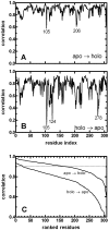
Figure 4. Analysis of ferric-binding protein.
A Upper panel displays the haemophilus influenzae ferric-binding protein in apo (purple; PDB code: 1D9V) and holo forms (green; PDB code: 1MRP). The two structures are superimposed on the fixed domain (residues 83–87, 102–225, 277–307). The Fe3+ ion is shown as a red sphere. Residues 9, 57, 175 and 193 are within 7 Å of the Fe atom. In addition, residues 8, 139–141, 176, 195 and 196 are in its 7–8 Å range. The lower panel shows the difference between the number of contacts of the ferric bound and unbound forms of FBP. B Contour map comparing the residue cross-correlations obtained from the detailed MD simulations of 10 ns duration (upper-right part of the map) and the coarse-grained model (lower-left part of the map) for holo-FBP. White and light gray are negative correlations, dark gray and black are positive correlations. Also displayed at the top of the figure are the detailed cross-correlations of residue 47 from the coarsened protein model (solid lines) and MD simulations (dashed lines). The value of zero is marked for each case to better discriminate positively and negatively correlated regions. The Pearson correlations between the data are 0.74 for the whole map and 0.94 for the selected residue, respectively.
Figure 5
Figure 5. Relative displacements of residues between the apo and holo forms (x-ray), and typical responses to a given force perturbation on residue 57 in the apo and holo forms.
The latter two curves are nudged for ease of comparison; their baselines are shown by dashed lines. Since the calculated displacement is proportional to the imposed force in LRT and therefore may be rescaled by a proportionality constant, the magnitude of the force is adjusted so as to make the average displacement the same as that of the x-ray experiment.
Figure 6
Figure 6. Displacement vectors between the perturbed and initial structures of ferric-binding protein.
The displacements are compared with those of the crystal structures, for A the apo, and B the holo form as the initial structure. In C, data in A and B are sorted from larges to smallest. Residues 47, 52, 130, 139–144, 147, 148, 166, 174, 186, 226, 232–236, 293, 296, 298, 299 give the highest and 105, 124, 278 the lowest correlations in the holo structure. The standard error on the mean increases with decreasing correlations, as determined from the averages of five perturbations in randomly selected directions. For correlations greater than 0.90, it is less than 0.05 for the apo, and 0.02 for the holo form; the largest errors on the mean are 0.2 for the least correlated values in both cases.
Figure 7
Figure 7. Remote modulation of Fe ion dissociation.
A The response of the protein to forces exerted on residue j = 47 in different directions (forces are shown with the red arrows.) The collection of all the displacements is shown in orange; responses on the three neighbors along the chain in either direction (44≤j≤50) are not shown for clarity. The volume taken up by the first neighbors of the Fe+3 is shaded. B The region of the first neighbors of the Fe+3 is magnified to display the response to perturbations applied at selected residues. All vector lengths are relative to the magnitude of the same size of unit response. Upper left figure magnifies the results in A. The Fe+3 ion contacting residues in the fixed domain (located on the right hand side) respond incoherently, moving mainly within a plane. Residues Q8, H9 and E57, that are in the moving domain of the protein respond coherently, moving parallel to each other, tending to open the cap for Fe ion to exit. Perturbations on regions in the binding domain (exemplified by E57 and Fe ion) also induce large changes in this region, the former in the moving domain, the latter in both domains, but coherence in cap residues no longer exists. Distant, non-controlling residues (e.g. V105) cannot induce large enough motions in this region, although coherence of the cap residues may exist (see Table 1). Figures are drawn with the VMD software .

References

    1. Zhang XJ, Wozniak JA, Matthews BW. Protein Flexibility and Adaptability Seen in 25 Crystal Forms of T4 Lysozyme. Journal of Molecular Biology. 1995;250:527–552. - PubMed
    1. Eisenmesser EZ, Millet O, Labeikovsky W, Korzhnev DM, Wolf-Watz M, et al. Intrinsic dynamics of an enzyme underlies catalysis. Nature. 2005;438:117–121. - PubMed
    1. Gunasekaran K, Ma BY, Nussinov R. Is allostery an intrinsic property of all dynamic proteins? Proteins-Structure Function and Bioinformatics. 2004;57:433–443. - PubMed
    1. Volkman BF, Lipson D, Wemmer DE, Kern D. Two-state allosteric behavior in a single-domain signaling protein. Science. 2001;291:2429–2433. - PubMed
    1. Tang C, Schwieters CD, Clore GM. Open-to-closed transition in apo maltose-binding protein observed by paramagnetic NMR. Nature. 2007;449:1078–1082. - PubMed

Publication types

MeSH terms