. 2009 Oct 23;4(10):e7583.
doi: 10.1371/journal.pone.0007583.

Critical role of activating transcription factor 4 in the anabolic actions of parathyroid hormone in bone

Affiliations

Critical role of activating transcription factor 4 in the anabolic actions of parathyroid hormone in bone

Shibing Yu et al. PLoS One. .

Abstract

Parathyroid hormone (PTH) is a potent anabolic agent for the treatment of osteoporosis. However, its mechanism of action in osteoblast and bone is not well understood. In this study, we show that the anabolic actions of PTH in bone are severely impaired in both growing and adult ovariectomized mice lacking bone-related activating transcription factor 4 (ATF4). Our study demonstrates that ATF4 deficiency suppresses PTH-stimulated osteoblast proliferation and survival and abolishes PTH-induced osteoblast differentiation, which, together, compromise the anabolic response. We further demonstrate that the PTH-dependent increase in osteoblast differentiation is correlated with ATF4-dependent up-regulation of Osterix. This regulation involves interactions of ATF4 with a specific enhancer sequence in the Osterix promoter. Furthermore, actions of PTH on Osterix require this same element and are associated with increased binding of ATF4 to chromatin. Taken together these experiments establish a fundamental role for ATF4 in the anabolic actions of PTH on the skeleton.


Conflict of interest statement

Competing Interests: The authors have declared that no competing interests exist.

Figures

Figure 1
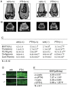
Figure 1. PTH-stimulated bone was significantly reduced or lost in Atf4−/− femurs.
A, two-dimensional (2D) reconstruction from μCT scan of femurs from growing wt, Atf4+/− and Atf4−/− mice treated with and without intermittent PTH for 28 d. B, quantitative analysis of bone volume/tissue volume (BV/TV), trabecular number (Tb. N), trabecular thickness (Tb.Th), trabecular space (Tb.Sp), and cortical thickness (Cort. Th). *P<0.05 (veh vs. PTH), P<0.05 (wt-veh vs. Atf4−/−-veh), #P<0.05 (PTH/veh-wt vs. PTH/veh-Atf4−/−).
Figure 2
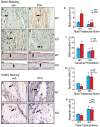
Figure 2. PTH-stimulated bone is severely impaired in Atf4−/− tibiae, vertebrae, and calvariae.
Representative H&E stained sections of tibiae (A–E), vertebrae (L5) (F–J), and calvariae (K–O) are shown. Trabecular bone area versus total area of tibiae (E) and vertebrae (J) was measured using an Image Pro Plus 6.2 software. The calvarial width was obtained from 20 random measurements throughout the whole calvaria using a SPOT Advanced imaging software (O). *P<0.05 (veh vs. PTH), P<0.05 (wt-veh vs. Atf4−/−-veh), #P<0.05 (PTH/veh-wt vs. PTH/veh-Atf4−/−).
Figure 3
Figure 3. Effects of ATF4 deficiency on PTH stimulation in adult OVX bone.
A, three-dimensional (3D) reconstruction from μCT scan of distal femurs of adult OVX mice. B, sagittal view of 2D distal femur at 1.7–2.0 mm from the chondro-osseous junction. C, BV/TV, Tb. N, Tb.Th, Tb.Sp, and Cort.Th. D, calcein double labeling of metaphyseal trabecular bone (magnification,x200). *P<0.05 (veh vs. PTH), P<0.05 (wt-veh vs. Atf4−/−-veh), #P<0.05 (PTH/veh-wt vs. PTH/veh-Atf4−/−).
Figure 4
Figure 4. Effects of PTH on osteoblast proliferation and survival in wt and Atf4−/− bone.
A–J, BrdU staining, sections of tibiae (A–D) and calvariae (F–I) were stained using a Zymed BrdU immunostaining kit. Proliferating cells were stained brown (arrows) and non-proliferating cells were stained blue. Proliferating cells on tibial trabecular surface or osteoid (E) or calvarial periosteal surface (J) were counted and normalized to total cells from the same area. K–P, TUNEL staining, sections of tibiae were stained using the ApopTag Peroxidase In Situ Apoptosis Detection Kit. Apoptotic osteoblasts and osteocytes (arrows) were stained brown and non-apoptotic cells were stained blue. Apoptotic osteoblasts and osteocytes in the trabecular (O) and cortical bone (P) of tibiae were counted and normalized to total osteoblasts and osteocytes from the same area. *P<0.05 (veh vs. PTH), P<0.05 (wt-veh vs. Atf4−/−-veh), #P<0.05 (PTH/veh-wt vs. PTH/veh-Atf4−/−).
Figure 5
Figure 5. Effects of PTH on expression of osteoblast marker genes in wt and ATF4 deficient mice.
A, quantitative real-time PCR, total RNAs were isolated from tibiae and analyzed by quantitative real-time RT-PCR using specific primers for Atf4, Ocn, Bsp, Col 1(I), ALP, Opn, Pthrp, c-Fos, and c-Jun mRNAs, which were normalized to Gapdh mRNA. *P<0.05 (veh vs. PTH), #P<0.05 (wt-veh vs. Atf4−/−-veh). B, plasma levels of IGF-1 and FGF-2 from mice using respective ELISA kits according to the manufacturer's instructions. *P<0.05 (veh vs. PTH),P<0.05 (wt-veh vs. Atf4−/−-veh), #P<0.05 (PTH/veh-wt vs. PTH/veh-Atf4−/−).
Figure 6
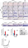
Figure 6. PTH fails to promote osteoblast maturation/differentiation in the absence of ATF4.
A–E, IHC analysis of Osx expression, sections of tibiae (A–D) and calvariae (E) were immunohistochemically stained using a specific antibody against Osx protein. The nuclei of Osx-positive cells (i.e., osteoblasts) were stained brown. The nuclei of preosteoblasts and other cells are stained blue. The total numbers of Osx-positive osteoblasts per tibial (G) or calvarial (H) section were counted under microscope. F, sections of tibiae were stained using an antibody against PTH1R protein. I, Western blot analysis, protein extracts were isolated from tibiae and analyzed for Osx, Runx2, and PTH1R proteins. *P<0.05 (veh vs. PTH), P<0.05 (wt-veh vs. Atf4−/−-veh), #P<0.05 (PTH/veh-wt vs. PTH/veh-Atf4−/−). J, cAMP assay, primary calvarial osteoblasts from 3-d-old wt or Atf4−/− mice were isolated, seeded at density of 5×10⁴ on 96-well plate, and treated with vehicle or increasing concentrations of human recombinant PTH(1-34) for 5 min followed by measurement of cAMP.
Figure 7
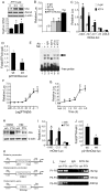
Figure 7. PTH activates Osx gene transcription via an ATF4-responsive element in the proximal Osx promoter.
A, MC-4 cells were electroporated with indicated amount of ATF4 expression plasmid followed by Western blot. B, COS-7 cells were transfected with p1060mOsx-luc, pRL-SV40, and indicated expression vectors followed by dual lucferase assays. C, COS-7 cells were transfected with various deletion constructs and pRL-SV40 with and without ATF4 expression plasmid. D, COS-7 cells transfected with p215mOsx-luc or the same plasmid containing a 3-bp substitution mutation in the putative ATF4-binding site and pRL-SV40 with and without ATF4 expression plasmid. E, EMSA, labeled wild-type DNA probe was incubated with 2 µg nuclear extracts from COS-7 cells transfected with pCMV/ATF4 plasmid in the presence of normal control IgG (lane 3), ATF4 antibody (lane 4), cFos antibody (lane 5), and ATF2 antibody (lane 6). Experiments were repeated 3–4 times and qualitatively identical results were obtained. F and G, MC-4 cells transfected with p1003mOsx-luc and pRL-SV40 were treated with indicated concentration of PTH for 6 h (F) or with 10−7 M PTH for indicated times (G). H, MC-4 cells were treated with and without 10−7 M PTH in the presence and absence of 10 µM of H89 for 6 h. I, MC-4 cells transfected as in Fig. 7C were treated with and without 10−7 M PTH for 6 h. J, MC-4 cells transfected as in Fig. D were treated with and without 10−7 M PTH for 6 h. K, a schematic illustration of putative ATF4 binding sites in the 5′ flanking regions of the Osx and osteocalcin gene promoters and osteocalcin gene. L, ChIP assay of the Osx promoter in MC-4 cells treated with and without 10−7 M PTH for 6 h. *P<0.05 (β-gal vs. ATF4, Runx2, and ATF4 plus Runx2, or veh vs. PTH), #P<0.05 (ATF4 plus Runx2 vs. β-gal, ATF4, or Runx2).
Figure 8
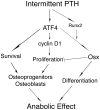
Figure 8. Proposed model for ATF4 mediation of PTH stimulation of bone formation.
Binding of PTH to PTH1R activates PKA and leads to up-regulation of ATF4. ATF4 subsequently increases proliferation and survival of osteoblasts. At the same time, ATF4 together with Runx2 maximally activates Osx expression and increases osteoblast differentiation. These increases in osteoblast number and differentiation lead to massive bone formation. Osx also negatively regulates osteoblast proliferation, thus preventing excess bone formation.

References

    1. Neer RM, Arnaud CD, Zanchetta JR, Prince R, Gaich GA, et al. Effect of parathyroid hormone (1-34) on fractures and bone mineral density in postmenopausal women with osteoporosis. N Engl J Med. 2001;344:1434–1441. - PubMed
    1. Miao D, He B, Karaplis AC, Goltzman D. Parathyroid hormone is essential for normal fetal bone formation. J Clin Invest. 2002;109:1173–1182. - PMC - PubMed
    1. Demiralp B, Chen HL, Koh AJ, Keller ET, McCauley LK. Anabolic actions of parathyroid hormone during bone growth are dependent on c-fos. Endocrinology. 2002;143:4038–4047. - PubMed
    1. Iida-Klein A, Zhou H, Lu SS, Levine LR, Ducayen-Knowles M, et al. Anabolic action of parathyroid hormone is skeletal site specific at the tissue and cellular levels in mice. J Bone Miner Res. 2002;17:808–816. - PubMed
    1. Iida-Klein A, Lu SS, Kapadia R, Burkhart M, Moreno A, et al. Short-term continuous infusion of human parathyroid hormone 1-34 fragment is catabolic with decreased trabecular connectivity density accompanied by hypercalcemia in C57BL/J6 mice. J Endocrinol. 2005;186:549–557. - PubMed

Publication types

LinkOut - more resources