

doi: 10.1007/s11011-009-9169-y.
Epub 2009 Oct 23.
Brain iron homeostasis, the choroid plexus, and localization of iron transport proteins
Affiliations
- PMID: 19851851
- PMCID: PMC2788140
- DOI: 10.1007/s11011-009-9169-y
Item in Clipboard
Brain iron homeostasis, the choroid plexus, and localization of iron transport proteins
Metab Brain Dis.
2009 Dec.
Abstract
Maintenance of appropriate iron homeostasis in the brain is important, but the mechanisms involved in brain iron uptake are incompletely understood. Here, we have analyzed where messenger RNAs that encode iron transport proteins are expressed in the brain, using the Allen Brain atlas, and we conclude that several important iron transporters are highly expressed in the choroid plexus. Based on recent estimates of the surface area of the choroid plexus and on MRI imaging studies of manganese uptake in the brain, we propose that the choroid plexus may have a much greater role than has been previously appreciated in brain iron transport.
Figures
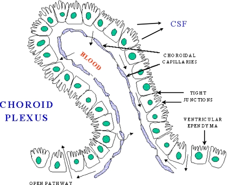
A schematic of the choroid plexus illustrates that the choroidal capillaries are separated from the CSF by a layer of choroidal epithelial cells. If the choroid plexus acted as a site of brain iron uptake, Tf released by the blood would bind to TfR on the membrane nearest to the capillary, and iron would cross to the membrane that faces the CSF, and be exported by FPN, aided by a ferroxidase such as ceruloplasmin or hephaestin. In this potential scheme, DMT1 would be needed to release iron from endosomes after internalization of the Tf-TfR complex, and an endosomal reductase would also be needed. This figure was reproduced from http://www.daviddarling.info/encyclopedia/copyright.html with permission from David Darling
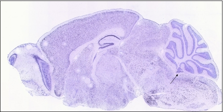
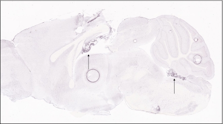
The transferrin receptor is expressed throughout the central nervous system, including in the choroid plexus, as judged by staining in a sagittal section of mouse brain from the Allen Mouse Brain atlas (arrow)
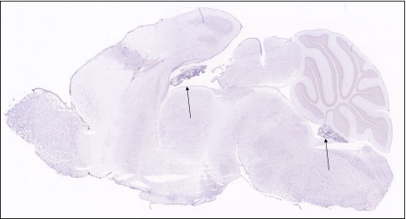
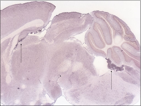
Expression of the divalent metal transporter, DMT1, is notably high in the choroid plexus, as indicated by arrows to choroid plexus formations
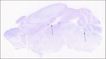
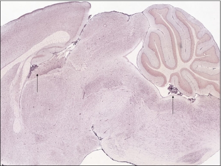
Expression of the reductase, Dcytb, is detectable in many regions of the brain, including in the choroid plexus (arrows)
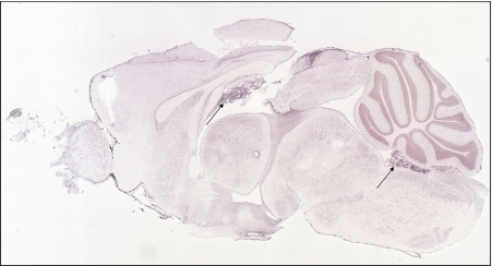
Ferroportin expression is very high in choroid plexus compared to other regions of the CNS (arrows)
Expression of the membrane-bound ferroxidase, hephaestin, is detectable throughout the brain, but is very high in the choroid plexus (arrows)
Expression of ceruloplasmin is also very high in the choroid plexus (arrows)
Expression of iron storage protein, ferritin H chain is highly expressed in the choroids plexus (arrows)
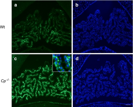
Immunofluorescent staining using anti-ferritin antibody (Dako) shows expression of ferritin in 24-months old mice choroid plexus (a). This immunoreactivity is highly increased in ceruloplasmin null mice (c) indicating increased intracellular iron in this area. DAPI staining b, d shows choroid plexus structure near fourth ventricle. Scale bars; 100 μm, 20 μm (inset)
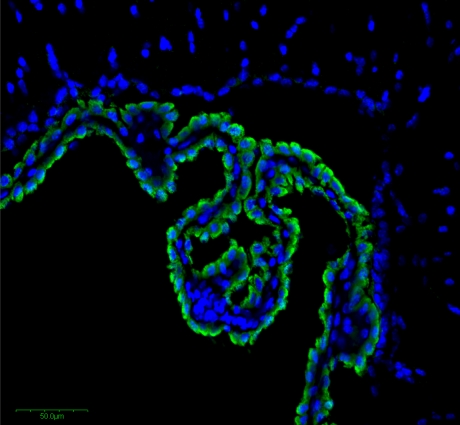
Immunohistochemistry reveals high expression of Dcytb in the choroid plexus epithelial cells, indicated by the green staining. Nuclei of epithelial, capillary and other cells are detected by staining with DAPI blue. Immunohistochemistry was performed on the frozen section of 10 μm thickness with a rabbit anti-mouse Dcytb polyclonal antibody as described previously (Su et al. ; Zhang et al. 2006)
Similar articles
-
Expression of copper trafficking genes in the mouse brain.Neuroreport. 1998 Oct 5;9(14):3259-63. doi: 10.1097/00001756-199810050-00023. Neuroreport. 1998. PMID: 9831461
-
Altered iron metabolism is part of the choroid plexus response to peripheral inflammation.Endocrinology. 2009 Jun;150(6):2822-8. doi: 10.1210/en.2008-1610. Epub 2009 Feb 12. Endocrinology. 2009. PMID: 19213835
-
Profile of altered brain iron acquisition in restless legs syndrome.Brain. 2011 Apr;134(Pt 4):959-68. doi: 10.1093/brain/awr012. Epub 2011 Mar 11. Brain. 2011. PMID: 21398376 Free PMC article.
-
Brain iron homeostasis.Dan Med Bull. 2002 Nov;49(4):279-301. Dan Med Bull. 2002. PMID: 12553165 Review.
-
Existing and emerging mechanisms for transport of iron and manganese to the brain.J Neurosci Res. 1999 Apr 15;56(2):113-22. doi: 10.1002/(SICI)1097-4547(19990415)56:2<113::AID-JNR1>3.0.CO;2-K. J Neurosci Res. 1999. PMID: 10777372 Review.
Cited by
-
H(+)-coupled divalent metal-ion transporter-1: functional properties, physiological roles and therapeutics.Curr Top Membr. 2012;70:169-214. doi: 10.1016/B978-0-12-394316-3.00005-3. Curr Top Membr. 2012. PMID: 23177986 Free PMC article. Review.
-
Brain Iron Metabolism Dysfunction in Parkinson's Disease.Mol Neurobiol. 2017 May;54(4):3078-3101. doi: 10.1007/s12035-016-9879-1. Epub 2016 Apr 2. Mol Neurobiol. 2017. PMID: 27039308 Review.
-
ATP7A-Regulated Enzyme Metalation and Trafficking in the Menkes Disease Puzzle.Biomedicines. 2021 Apr 6;9(4):391. doi: 10.3390/biomedicines9040391. Biomedicines. 2021. PMID: 33917579 Free PMC article. Review.
-
Gene expression and functional annotation of the human and mouse choroid plexus epithelium.PLoS One. 2013 Dec 31;8(12):e83345. doi: 10.1371/journal.pone.0083345. eCollection 2013. PLoS One. 2013. PMID: 24391755 Free PMC article.
-
The Aging of Iron Man.Front Aging Neurosci. 2018 Mar 12;10:65. doi: 10.3389/fnagi.2018.00065. eCollection 2018. Front Aging Neurosci. 2018. PMID: 29593525 Free PMC article. Review.
References
-
- Beard JL, Hendricks MK, Perez EM, Murray-Kolb LE, Berg A, Vernon-Feagans L, Irlam J, Isaacs W, Sive A, Tomlinson M. Maternal iron deficiency anemia affects postpartum emotions and cognition. J Nutr. 2005;135:267–272. - PubMed
Publication types
MeSH terms
Substances
Grants and funding
LinkOut - more resources
Full Text Sources
Medical
