

Epithelial-mesenchymal transition in hepatocellular carcinoma
- PMID: 19852728
- PMCID: PMC2963061
- DOI: 10.2217/fon.09.91
Epithelial-mesenchymal transition in hepatocellular carcinoma
Abstract
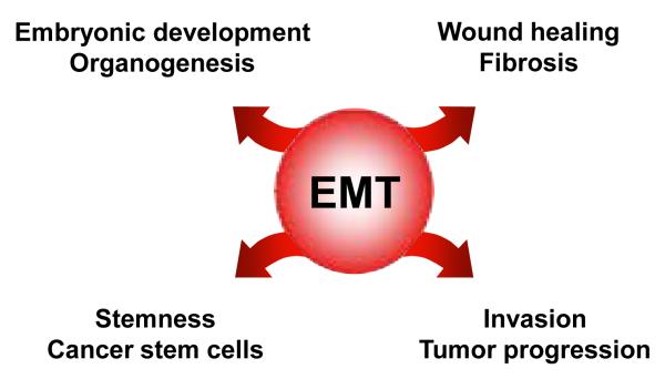
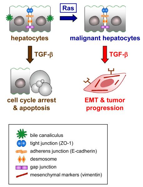
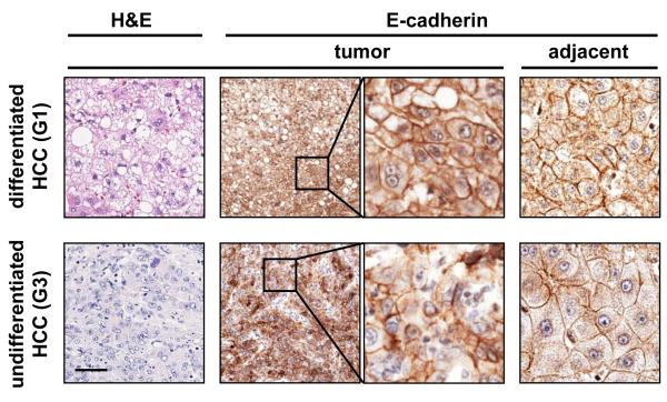
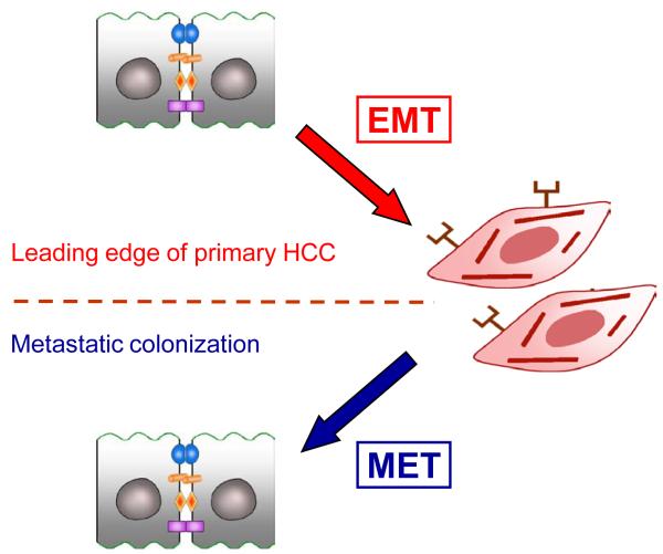
The transition of epithelial cells to a mesenchymal phenotype is of paramount relevance for embryonic development and adult wound healing. During the past decade, the epithelial-mesenchymal transition (EMT) has been increasingly recognized to occur during the progression of various carcinomas such as hepatocellular carcinoma (HCC). Here, we focus on EMT in both experimental liver models and human HCC, emphasizing the underlying molecular mechanisms which show partial recurrence of embryonic programs such as TGF-beta and Wnt/ beta-catenin signaling, including collaboration with hepatitis viruses. We further discuss the differentiation repertoire of malignant hepatocytes with respect to the potential acquisition of stemness, and the involvement of the mesenchymal to epithelial transition, the reversal of EMT, in cancer dissemination and metastatic colonization. The strong evidence for EMT in HCC patients demands novel strategies in pathological assessments and therapeutic concepts to efficiently combat HCC progression.
Figures
References
-
- El-Serag HB, Rudolph KL. Hepatocellular carcinoma: epidemiology and molecular carcinogenesis. Gastroenterology. 2007;132:2557–2776. - PubMed
-
- Kensler TW, Qian GS, Chen JG, Groopman JD. Translational strategies for cancer prevention in liver. Nat Rev Cancer. 2003;3:321–329. - PubMed
-
- Farazi PA, DePinho RA. Hepatocellular carcinoma pathogenesis: from genes to environment. Nat Rev Cancer. 2006;6:674–687. - PubMed
-
- Jou J, Choi SS, Diehl AM. Mechanisms of disease progression in nonalcoholic fatty liver disease. Semin Liver Dis. 2008;28:370–379. - PubMed
Publication types
MeSH terms
Grants and funding
LinkOut - more resources
Full Text Sources
Other Literature Sources
Medical
Miscellaneous