. 2009 Oct 23:4:47.
doi: 10.1186/1748-717X-4-47.

AT-101, a small molecule inhibitor of anti-apoptotic Bcl-2 family members, activates the SAPK/JNK pathway and enhances radiation-induced apoptosis

Affiliations

AT-101, a small molecule inhibitor of anti-apoptotic Bcl-2 family members, activates the SAPK/JNK pathway and enhances radiation-induced apoptosis

Shuraila F Zerp et al. Radiat Oncol. .

Abstract

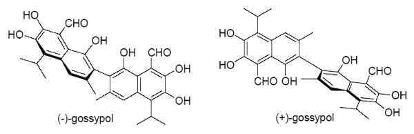
Background: Gossypol, a naturally occurring polyphenolic compound has been identified as a small molecule inhibitor of anti-apoptotic Bcl-2 family proteins. It induces apoptosis in a wide range of tumor cell lines and enhances chemotherapy- and radiation-induced cytotoxicity both in vitro and in vivo. Bcl-2 and related proteins are important inhibitors of apoptosis and frequently overexpressed in human tumors. Increased levels of these proteins confer radio- and chemoresistance and may be associated with poor prognosis. Consequently, inhibition of the anti-apoptotic functions of Bcl-2 family members represents a promising strategy to overcome resistance to anticancer therapies.

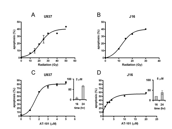
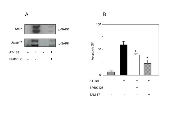
Methods: We tested the effect of (-)-gossypol, also denominated as AT-101, radiation and the combination of both on apoptosis induction in human leukemic cells, Jurkat T and U937. Because activation of the SAPK/JNK pathway is important for apoptosis induction by many different stress stimuli, and Bcl-X(L) is known to inhibit activation of SAPK/JNK, we also investigated the role of this signaling cascade in AT-101-induced apoptosis using a pharmacologic and genetic approach.

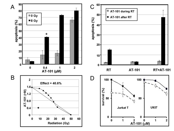
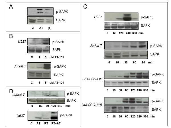
Results: AT-101 induced apoptosis in a time- and dose-dependent fashion, with ED50 values of 1.9 and 2.4 microM in Jurkat T and U937 cells, respectively. Isobolographic analysis revealed a synergistic interaction between AT-101 and radiation, which also appeared to be sequence-dependent. Like radiation, AT-101 activated SAPK/JNK which was blocked by the kinase inhibitor SP600125. In cells overexpressing a dominant-negative mutant of c-Jun, AT-101-induced apoptosis was significantly reduced.

Conclusion: Our data show that AT-101 strongly enhances radiation-induced apoptosis in human leukemic cells and indicate a requirement for the SAPK/JNK pathway in AT-101-induced apoptosis. This type of apoptosis modulation may overcome treatment resistance and lead to the development of new effective combination therapies.


Figures

Figure 1
Figure 1
Chemical structure of the (-) and (+) enantiomer of gossypol.
Figure 2
Figure 2
Dose-dependent induction of apoptosis by radiation (A, B) and AT-101 (C, D) in human leukemic U937 (A, C) and Jurkat T cells (B, D). Apoptosis was quantified by FACScan analysis at t = 24 h after treatment. Data are presented as mean values (± SD) from 3 independent experiments. Inserts in C and D show the time-dependency of AT-101.
Figure 3
Figure 3
Synergistic and sequence-dependent interaction between radiation and AT-101 in U937 cells. A: The combination of radiation and AT-101 induces more apoptosis than the sum of the effects caused by the single agent treatment. Hatched bars represent the apoptotic effect by AT-101 alone (0-2 μM); black bars represent the combined effect with radiation (8 Gy). B: Isobolographic analysis of the combined effect of 40.6% apoptosis (* in A) induced by 0.4 μM AT-101 and 8 Gy radiation. The combination point is projected below the area of additivity, indicating synergy. The combination index for this point: CI = 0.42. C: Sequence-dependency of radiation and AT-101. Radiation (6 Gy) and AT-101 (1 μM) were either applied concurrently (hatched bars) or sequentially (AT-101 24 h after radiation; black bars). Apoptosis was analyzed at t = 24 h after AT-101. D: MTT cell viability assays in Jurkat T and U937 cells. AT-101 was added at the indicated concentrations (solid lines); radiation was dosed at 8 Gy (dashed line). Viability was determined at t = 48 h after radiation (i.e. 24 h after AT-101). Data presented in A, C and D are mean values (± SD) from 2 independent experiments.
Figure 4
Figure 4
Gossypol and radiation activate the SAPK/JNK pathway. A: AT-101 is a stronger activator of SAPK/JNK than racemic (±)-gossypol. U937 cells were treated with equimolar concentrations of AT-101 (5 μM) and SAPK/JNK activation was analyzed at t = 2 h. (Abbreviations: C = control; AT = AT-101; ± =(±)-gossypol). B: Dose-dependent SAPK/JNK activation in U937 (upper panel) and Jurkat T cells (lower panel). Cells were treated with indicated concentrations of AT-101 and SAPK/JNK activation was analyzed at t = 2 h. C: Kinetics of 5 μM AT-101-induced SAPK/JNK in human leukemic (U937 and Jurkat T) and carcinoma cells (VU-SCC-OE and UM-SCC-11B). D: Radiation (8 Gy) induces a time-dependent SAPK/JNK activation in Jurkat T cells (upper panel). In U937 cells, the combination of AT-101 (AT; 5 μM) and radiation (RT; 10 Gy) induces a stronger activation of SAPK/JNK at t = 2 h than single modality treatment (lower panel).
Figure 5
Figure 5
AT-101 employs the SAPK/JNK pathway to induce apoptosis. A: AT-101 (5 μM) induced SAPK/JNK in U937 and Jurkat T cells can be inhibited by the SP600125 kinase inhibitor; t = 90 min. B: Blockade of SAPK/JNK signaling by kinase inhibitor (SP600125) or dominant-negative c-Jun (TAM-67) inhibits AT-101 (5 μM)-induced apoptosis at t = 20 h in U937 cells. Data are presented as mean values (± SD) from 2 independent experiments. *p < 0.005, Student's t test.

Similar articles

Cited by

References

    1. Belka C, Jendrossek V, Pruschy M, Vink S, Verheij M, Budach W. Apoptosis-modulating agents in combination with radiotherapy-current status and outlook. Int J Radiat Oncol Biol Phys. 2004;58:542–554. - PubMed
    1. Hellemans P, van Dam PA, Weyler J, Van Oosterom AT, Buytaert P, Van Marck E. Prognostic value of bcl-2 expression in invasive breast cancer. Br J Cancer. 1995;72:354–360. - PMC - PubMed
    1. Krajewski S, Krajewska M, Ehrmann J, Sikorska M, Lach B, Chatten J, Reed JC. Immunohistochemical analysis of Bcl-2, Bcl-X, Mcl-1, and Bax in tumors of central and peripheral nervous system origin. Am J Pathol. 1997;150:805–814. - PMC - PubMed
    1. Olopade OI, Adeyanju MO, Safa AR, Hagos F, Mick R, Thompson CB, Recant WM. Overexpression of BCL-x protein in primary breast cancer is associated with high tumor grade and nodal metastases. Cancer J Sci Am. 1997;3:230–237. - PubMed
    1. Pena JC, Thompson CB, Recant W, Vokes EE, Rudin CM. Bcl-xL and Bcl-2 expression in squamous cell carcinoma of the head and neck. Cancer. 1999;85:164–170. doi: 10.1002/(SICI)1097-0142(19990101)85:1<164::AID-CNCR23>3.0.CO;2-Q. - DOI - PubMed

Publication types

MeSH terms

Substances