. 2010 Feb;31(5):964-74.
doi: 10.1016/j.biomaterials.2009.10.010. Epub 2009 Oct 22.

Doxycycline loaded poly(ethylene glycol) hydrogels for healing vesicant-induced ocular wounds

Affiliations

Doxycycline loaded poly(ethylene glycol) hydrogels for healing vesicant-induced ocular wounds

SivaNaga S Anumolu et al. Biomaterials. 2010 Feb.

Abstract

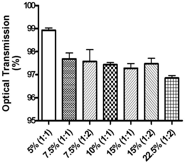
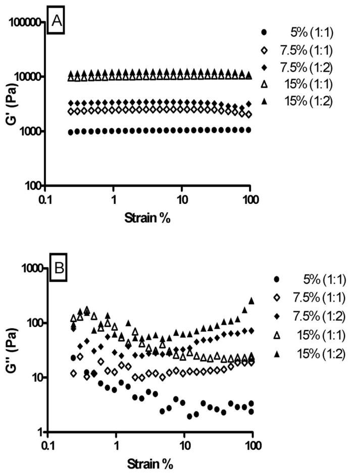
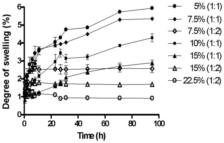
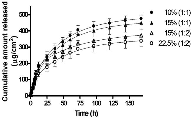
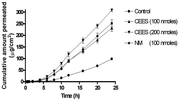
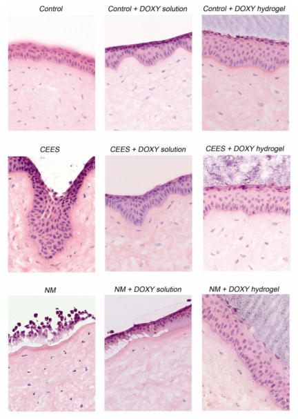
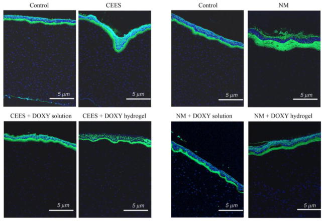
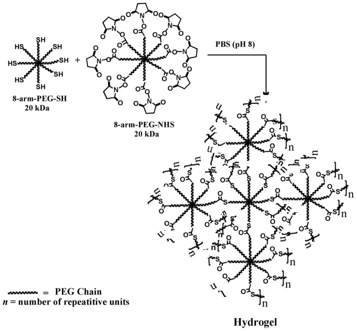
Half mustard (CEES) and nitrogen mustard (NM) are commonly used surrogates and vesicant analogs of the chemical warfare agent sulfur mustard. In the current study, in situ forming poly(ethylene glycol) (PEG)-based doxycycline hydrogels are developed and evaluated for their wound healing efficacy in CEES and NM-exposed rabbit corneas in organ culture. The hydrogels, characterized by UV-Vis spectrophotometry, rheometry, and swelling kinetics, showed that the hydrogels are optically transparent, have good mechanical strength and a relatively low degree of swelling (<7%). In vitro doxycycline release from the hydrogel disks (0.25% w/v) was found to be biphasic with release half times of approximately 12 and 72h, respectively, with 80-100% released over a 7-day period. Permeation of doxycycline through vesicant wounded corneas was found to be 2.5 to 3.4 fold higher than non-wounded corneas. Histology and immunofluorescence studies showed a significant reduction of matrix metalloproteinase-9 (MMP-9) and improved healing of vesicant-exposed corneas by doxycycline hydrogels compared to a similar dose of doxycycline delivered in phosphate buffered saline (PBS, pH 7.4). In conclusion, the current studies demonstrate that the doxycycline-PEG hydrogels accelerate corneal wound healing after vesicant injury offering a therapeutic option for ocular mustard injuries.


Figures

Fig. 1
Fig. 1
Optical transmission of 5% (1:1), 7.5% (1:1), 7.5% (1:2), 10% (1:1), 15% (1:2), 15% (1:1), and 22.5% (1:2) hydrogels. All the hydrogels were found to be transparent. A change in hydrogel composition resulted in a statistically significant effect (p<0.05) on their optical transmission properties. A slight decrease in transparency of the hydrogels was observed with an increase in the polymer concentration and their crosslinking ratios.
Fig. 2
Fig. 2
Influence of strain on G′ (A) and G″ (B) of 5% (1:1), 7.5% (1:1), 7.5% (1:2), 15% (1:1), and 15% (1:2) hydrogels. The strain sweep test establishes the range of linear viscoelasticity (LVE) for the hydrogels.
Fig. 3
Fig. 3
Influence of frequency on G′ (A) and G″ (B) of 5% (1:1), 7.5% (1:1), 7.5% (1:2), 15% (1:1), and 15% (1:2) hydrogels. The frequency sweep test shows that the hydrogels are more elastic than viscous and that they have the ability to resist structural changes under strain. A change in hydrogel composition resulted in a statistically significant effect (p<0.001) on the viscoelasticity of the hydrogels.
Fig. 4
Fig. 4
Effect of polymer concentration and crosslinking density on the swelling kinetics of 5% (1:1), 7.5% (1:1), 7.5% (1:2), 10% (1:1), 15% (1:1), 15% (1:2), and 22.5% (1:2) hydrogels. The higher the concentration of polymers and crosslinking density, the lower is the degree of swelling.
Fig. 5
Fig. 5
Cumulative amount of doxycycline released as a function of time for hydrogels: 10% (1:1), 15% (1:2), 15% (1:1), and 22.5% (1:2). The release data were fitted using a two-phase exponential association equation in GraphPad Prism 4 software. The goodness of fit varied from 0.87 to 0.99. The release mechanism is non-Fickian or anomalous involving both diffusion and polymer relaxation (0.5
Fig. 6
Fig. 6
Cumulative amount of doxycycline permeated as a function of time through cornea exposed to different concentrations of CEES and NM. The permeability of doxycycline through CEES and NM-exposed corneas was significantly higher than the controls (p<0.0001) by 2.5 to 3.3 fold.
Fig. 7
Fig. 7
H & E staining to visualize the histology of CEES and NM-exposed corneas treated for 24 h with doxycycline in solution or in a hydrogel. The damaged area is where the epithelium meets the stroma. The wound healing efficacy of doxycycline solution was close to the doxycycline hydrogel for CEES exposed corneas, as the extent of damage was comparatively mild. However, a superior wound healing efficacy was observed with hydrogels over solutions when harshly damaged NM-exposed corneas were treated with doxycycline.
Fig. 8
Fig. 8
Immunofluorescent staining of corneas exposed to CEES and NM and subsequently treated with doxycycline either in solution or hydrogel. The intensity of MMP-9 staining, increased from exposure to CEES and NM, was significantly decreased by doxycycline both in solution and hydrogel. However, the doxycycline hydrogel also improved the attachment between the epithelium and stroma after NM exposure. This demonstrated that the doxycycline hydrogel formulation was more effective than doxycycline in solution.
Scheme 1
Scheme 1
Hydrogel formation by intermolecular crosslinking of PEG polymers containing mutually reactive thiol (8-arm-PEG-SH) and N-hydroxysuccinimidyl ester (8-arm-PEG-NHS) groups at room temperature in buffer (pH 8). The hydrogel networks are produced by the formation of thioester bonds.
Scheme 2
Scheme 2
Mechanism of formation of thioester bonds by reaction of thiol group with N-hydroxysuccinimidyl ester. The 8-arm-PEG-SH (I) exists as a thiolate (II) in PBS (pH 8), which acts as a nucleophile and attacks the carbonyl carbon of 8-arm-PEG-NHS (III) to form the intermediate (IV) leading to the formation of thioester bonds accompanied by the cleavage of N-hydroxysuccinimide.

Similar articles

Cited by

References

    1. Blanc PD. The legacy of war gas. Am J Med. 1999;106(6):689–690. - PubMed
    1. Saladi RN, Smith E, Persaud AN. Mustard: a potential agent of chemical warfare and terrorism. Clin Exp Dermatol. 2006;31(1):1–5. - PubMed
    1. Borak J, Sidell FR. Agents of chemical warfare: sulfur mustard. Ann Emerg Med. 1992;21(3):303–308. - PubMed
    1. Ghanei M, Harandi AA. Long term consequences from exposure to sulfur mustard: a review. Inhal Toxicol. 2007;19(5):451–456. - PubMed
    1. Papirmeister B. In: Medical Defense Against Mustard Gas: Toxic Mechanisms and Pharmacological Implications. Papirmeister B, Feister AJ, Robinson SI, Ford RD, editors. Boca Raton, Fl: CRC Press; 1991.

Publication types