. 2009 Dec 15;147(1-3):265-76.
doi: 10.1016/j.pain.2009.09.030. Epub 2009 Oct 22.

Peripheral and central sensitization in remote spinal cord regions contribute to central neuropathic pain after spinal cord injury

Affiliations

Peripheral and central sensitization in remote spinal cord regions contribute to central neuropathic pain after spinal cord injury

Susan M Carlton et al. Pain. .

Abstract

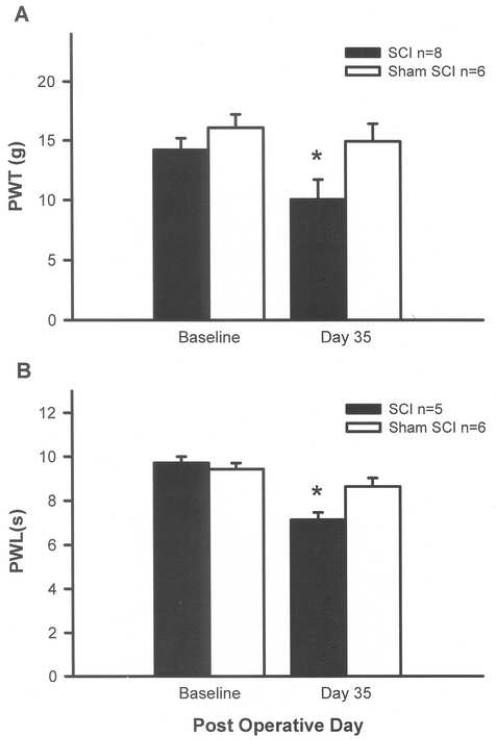
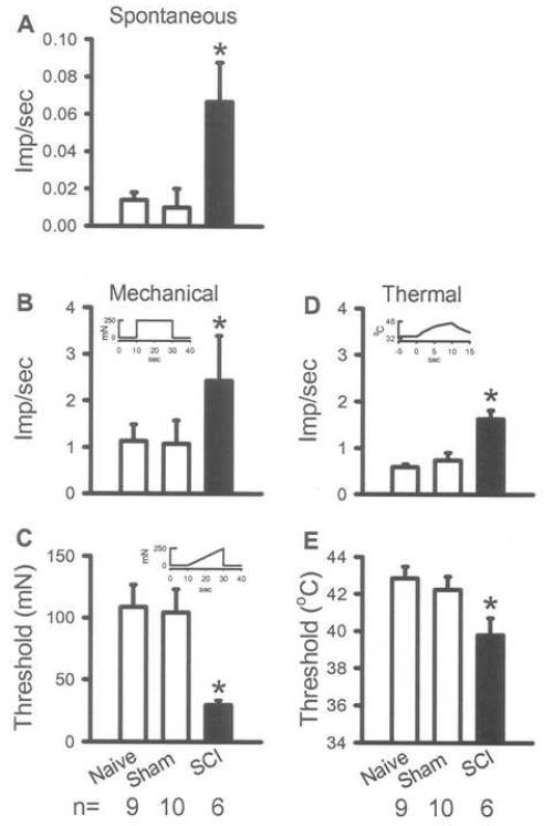
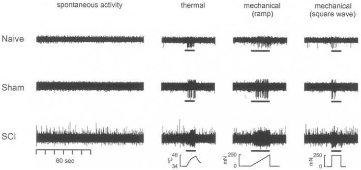
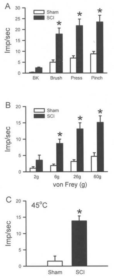
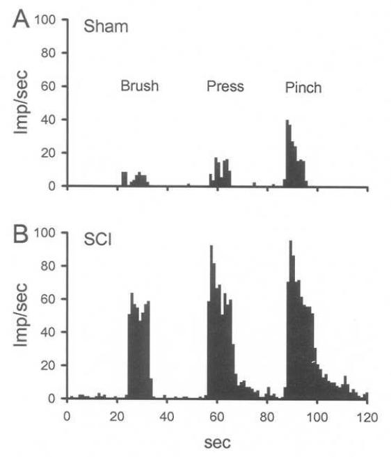
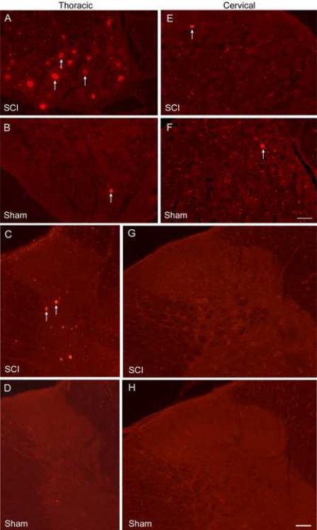
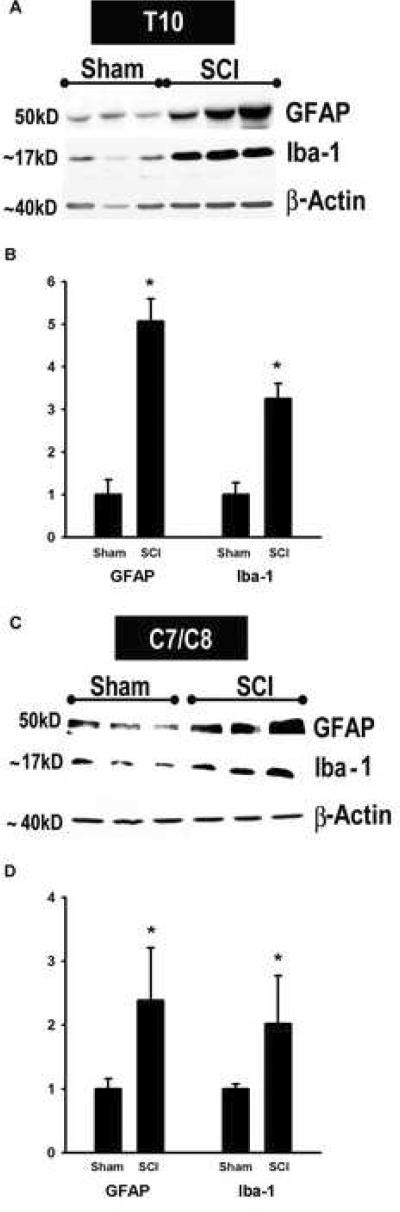
Central neuropathic pain (CNP) developing after spinal cord injury (SCI) is described by the region affected: above-level, at-level and below-level pain occurs in dermatomes rostral, at/near, or below the SCI level, respectively. People with SCI and rodent models of SCI develop above-level pain characterized by mechanical allodynia and thermal hyperalgesia. Mechanisms underlying this pain are unknown and the goals of this study were to elucidate components contributing to the generation of above-level CNP. Following a thoracic (T10) contusion, forelimb nociceptors had enhanced spontaneous activity and were sensitized to mechanical and thermal stimulation of the forepaws 35 days post-injury. Cervical dorsal horn neurons showed enhanced responses to non-noxious and noxious mechanical stimulation as well as thermal stimulation of receptive fields. Immunostaining dorsal root ganglion (DRG) cells and cord segments with activating transcription factor 3 (ATF3, a marker for neuronal injury) ruled out neuronal damage as a cause for above-level sensitization since few C8 DRG cells expressed AFT3 and cervical cord segments had few to no ATF3-labeled cells. Finally, activated microglia and astrocytes were present in thoracic and cervical cord at 35 days post-SCI, indicating a rostral spread of glial activation from the injury site. Based on these data, we conclude that peripheral and central sensitization as well as reactive glia in the uninjured cervical cord contribute to CNP. We hypothesize that reactive glia in the cervical cord release pro-inflammatory substances which drive chronic CNP. Thus a complex cascade of events spanning many cord segments underlies above-level CNP.


Figures

Fig. 1
Fig. 1
SCI effects forelimb sensitivity. At 35 days post-contusion, SCI animals have mechanical allodynia evidenced by a decrease in paw withdrawal threshold (PWT) to von Frey stimulation (A) and have thermal hyperalgesia evidenced by a decrease in paw withdrawal latency (PWL) to thermal stimulation (B). Sham rats show no change from baseline. *p < 0.05, repeated measures ANOVA.
Fig. 2
Fig. 2
Peripheral sensitization of forelimb primary afferents 35 days following SCI. In vitro recordings from nociceptors demonstrate an increased spontaneous activity compared to naïve and sham animals (A). These units also demonstrate mechanical sensitization evidenced by an increased response to a suprathreshold mechanical stimulation (B) and a decreased threshold to activation (C). Units demonstrate thermal hyperalgesia evidenced by an increased response to thermal stimulation and a decreased threshold to activation (E). Insets in B, C and D show stimulus parameters. *p<0.05, Kruskal-Wallis test followed by a Dunn's post hoc analysis.
Fig. 3
Fig. 3
Recordings from nociceptors in SCI rats show sensitization. At 35 days post-injury, spontaneous activity in nociceptors is clearly enhanced in SCI compared to naïve and sham animals. Evoked responses following thermal and mechanical stimulation are also greater in magnitude in SCI compared to naïve and sham animals. (Data is unfiltered; stimulus is shown at the bottom of each column.)
Fig. 4
Fig. 4
Sensitization of responses of dorsal horn neurons 35 days following SCI. Recordings from unidentified dorsal horn neurons demonstrate significantly enhanced responses to brush, press and pinch in the receptive fields of the neurons. Responses to the von Frey filaments and thermal stimulation are also significantly enhanced. *p<0.05, one way ANOVA followed by a Tukey's test.
Fig. 5
Fig. 5
Recordings from dorsal horn neurons in SCI rats show sensitization. Peristimulus histograms show that compared to sham (A), cervical WDR neurons in SCI rats (B) are sensitized evidenced by the enhanced responses to brush, press and pinch in their receptive fields.
Fig. 6
Fig. 6
ATF3 immunostaining in DRG and spinal cord. At 2 days post-contusion, the T10 DRG has numerous ATF3-stained nuclei (A); however, the sham T10 DRG has few to none (B). The spinal cord near T10 in SCI rats has many ATF3-labeled neurons (C) but the thoracic cord in sham rats has none (D). In the cervical region, a very small number of DRG cells from SCI (E) or sham (F) rats are labeled for ATF3. Similarly, in the cervical cord in SCI (G) or sham (H) rats, almost no ATF3 cells are observed. White arrows indicate ATF3-stained nuclei. Bar = 50μm
Fig. 7
Fig. 7
ATF3 labels neurons. Sections from the T9 DRG (A) and spinal cord (B) 2 days post-injury were double labeled for ATF3 (green) and NeuN (red, a neuronal marker) to confirm neuronal, and not glial, expression of ATF3. All AFT3-expressing cells also stained for NeuN (arrows). The asterisk identifies a single labeled NeuN cell. Bar = 25μm
Fig. 8
Fig. 8
Spread of glial activation in chronically injured spinal cords. Using Western blots, GFAP (marker of astrocytic activation, ~ 50kD) and Iba-1 (marker of microglial activation, ~17kD) levels were measured at the site of injury (T10) and in cervical segments (C7/C9), 35 days post-injury (A and C). n = 3 sham and 3 SCI. Quantitative analyses of immunoblotted GFAP and Iba-1 expression in T10 (B, n = 6 sham, 9 SCI) and C7/C8 (D, n = 3 sham, 3 SCI). The Y-axis represents the relative intensity of the GFAP and Iba-1 bands normalized to β-actin and then to sham values (set to 1). GFAP and Iba-1 protein levels show significant increases 35d after SCI at both the site of injury and in the cervical cord. *p<0.05, Student's t-test.

References

    1. Basso DM, Beattie MS, Bresnahan JC. A sensitive and reliable locomotor rating scale for open field testing in rats. J Neurotrauma. 1995;121:1–21. - PubMed
    1. Bennett AD, Everhart AW, Hulsebosch CE. Intrathecal administration of an NMDA or a non-NMDA receptor antagonist reduces mechanical but not thermal allodynia in a rodent model of chronic central pain after spinal cord injury. Brain Res. 2000;8591:72–82. - PubMed
    1. Bong GW, Rosengren S, Firestein GS. Spinal cord adenosine receptor stimulation in rats inhibits peripheral neutrophil accumulation. The role of N-methyl-D-aspartate receptors. J Clin Invest. 1996;9812:2779–2785. - PMC - PubMed
    1. Bontioti E, Dahlin LB, Kataoka K, Kanje M. End-to-side nerve repair induces nuclear translocation of activating transcription factor 3. Scand J Plast Reconstr Surg Hand Surg. 2006;406:321–328. - PubMed
    1. Boyle DL, Moore J, Yang L, Sorkin LS, Firestein GS. Spinal adenosine receptor activation inhibits inflammation and joint destruction in rat adjuvant-induced arthritis. Arthritis Rheum. 2002;4611:3076–3082. - PubMed

Publication types

MeSH terms

Substances