

Review
doi: 10.1016/j.devcel.2009.09.011.
Arrestin development: emerging roles for beta-arrestins in developmental signaling pathways
Affiliations
- PMID: 19853559
- PMCID: PMC3221601
- DOI: 10.1016/j.devcel.2009.09.011
Item in Clipboard
Review
Arrestin development: emerging roles for beta-arrestins in developmental signaling pathways
Dev Cell.
2009 Oct.
Abstract
Arrestins were identified as mediators of G protein-coupled receptor (GPCR) desensitization and endocytosis. However, it is now clear that they scaffold many intracellular signaling networks to modulate the strength and duration of signaling by diverse types of receptors--including those relevant to the Hedgehog, Wnt, Notch, and TGFbeta pathways--and downstream kinases such as the MAPK and Akt/PI3K cascades. The involvement of arrestins in many discrete developmental signaling events suggests an indispensable role for these multifaceted molecular scaffolds.
Figures

(1) Upon Hh binding, Ptc-mediated repression of Smo is released, resulting in (2) phosphorylation of Smo by GRK-2 and formation of a complex between Smo, β-arrestins, and the molecular motor Kif3A. (3) The Smo-β-arrestin-Kif3A complex translocates Smo to the primary cilium where (4) Smo cleaves Gli into its active form. (5) Active Gli then translocates down the primary cilium and (6) into the nucleus where it activates transcription of downstream targets.

(1) In canonical Wnt signaling, in the absence of receptor stimulation, the Axin-APC complex degrades β-catenin and strongly represses transcriptional activity. (2) Upon Wnt binding to Fz, β-arrestins bind to the receptor through Dsh, thus sequestering Axin and GSK3 away from β-catenin, (3) leading to its stabilization. (4) β-arrestins also act to internalize Fzs into endosomes while (5) β-catenin is free to translocate into the nucleus and initiate transcription. (6) During noncanonical Wnt signaling, β-arrestin complexes with Dsh and AP-2 after Wnt binding to Fz. (7) Subsequently, β-arrestin activates the GTPases RhoA and Rac1 leading to (8) ROCK activation, (9) actin reorganization and convergent extension, or (10) JNK activation.

(1) Upon ligand binding to the TβRI, -RII, and -RIII type receptors, (2) β-arrestin binds TβRIII (3) while TbRII phophorylates TβRI, which in turn activates R-Smads. (4) Activated R-Smads complex with co-Smads and (5) translocate into the nucleus where they initiate transcription. (6) Meanwhile, β-arrestin internalizes TβRIII, (7) thus attenuating TGFβ-mediated activation of R-Smads. (8) Additionally, TβRIII is able to activate p38 in a Smad-independent manner after TGFβ binding and (9) β-arrestin-mediated internalization of TβRIII attenuates this signaling as well. (10) Downstream of TGFβ-binding, β-arrestin is essential for the activation of Cdc42, which is responsible for (11) actin reorganization leading to (12) chemotaxis and filipodial extension.
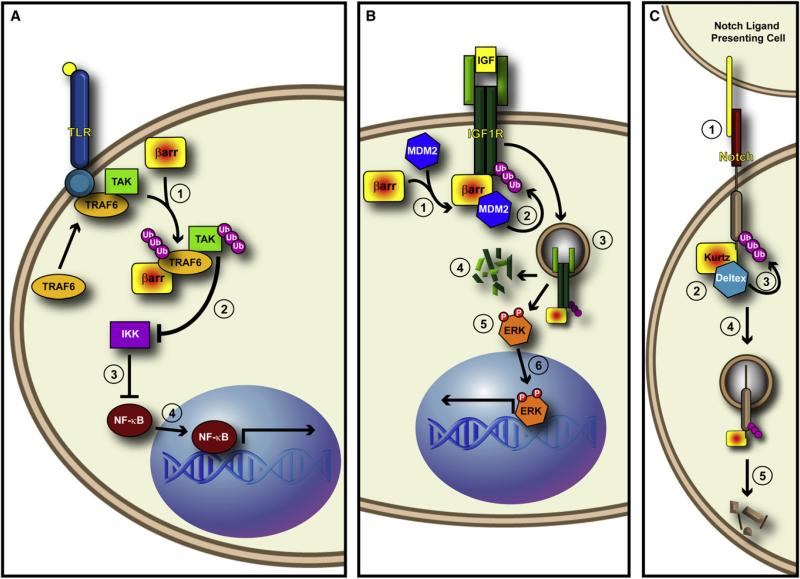
(A) β-arrestin promotes the ubiquitination of TRAF6. (1) Upon binding of ligand to the Toll-like Receptor, β-arrestin binds to and promotes the autoubiquitination of TRAF6. This autoubiquitination event is required for downstream activation of TAK and subsequent activation of NF-κB signaling. (2) It may be that β-arrestin-mediated ubiquitination of TRAF6 leads to activation of TAK, which inhibits IKK, (3) thus freeing NF-κB from IKK-mediated inhibition. (4) This would then allow NF-kB translocation into the nucleus and activation of transcription. (B) β-arrestin acts as an E3 ligase adaptor in response to IGF stimulation. (1) After IGF binds to the tetrameric IGF1R, β-arrestin recruits Mdm2 to the receptor. (2) Mdm2 ubiquitinates IGF1R, thus (3) leading to its internalization. (4) Once internalized, IGF1R is degraded by the protoesome, and (5) β-arrestin mediates the activation of ERK from internalized “signalosomes.” (6) ERK then translocates to the nucleus and activates transcription. (C) Krz mediates the ubiquitination and degradation of Drosophila Notch. (1) A Notch ligand on the Notch-ligand presenting cell binds Notch. (2) This event triggers the formation of a complex between Krz and the E3 ligase Dx. (3) Krz brings Dx to Notch and promotes Notch ubiquitination, (4) which leads to the Krz-dependent internalization of Notch, and (5) its subsequent degradation.
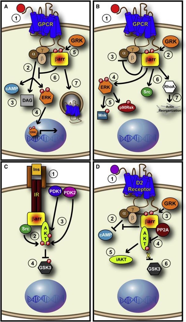
(A) GPCRs generate rapid and transient ERK, which is attenuated by β-arrestins. (1) As ligand (red circle) binds to the extracellular domain of a GPCR, G proteins are activated (2) and generate second messengers such as cAMP and DAG, while (3) also stimulating the rapid and transient phosphorylation of ERK. (4) This “G protein-stimulated” ERK translocates into the nucleus and modulates transcription. Meanwhile, (5) GRKs phosphorylate the C-terminal tail of receptors, thus (6) recruiting β-arrestins that sterically inhibit G protein binding, thus attenuating generation of second messengers. (7) β-arrestins also assist in the internalization of receptors, thus leading to their downregulation. (B) β-arrestins act as signaling adaptors and activate cytosolic ERK in a persistent fashion. (1) Upon ligand binding, (2) β-arrestins are recruited to the receptor and (3) block G protein-mediated signaling. (4) β-arrestins are able to stimulate the phosphorylation of ERK, which leads to a slowly developing, persistent pool of “β-arrestin-stimulated” ERK that resides exclusively in the cytosol and phosphorylates cytoplasmic substrates such as (5) Mnk and p90Rsk. (6) β-arrestins are also able to stimulate proteins such as c-Src and (7) RhoA, leading to a wide range of responses. (C) β-arrestin positively regulates Akt in response to Insulin Receptor Stimulation. (1) Bindng of Insulin (Ins) to the Insulin Receptor (IR) triggers the binding of β-arrestin 2 to the receptor. (2) β-arrestin scaffolds c-Src to Akt, causing the phosphorylation of Akt by c-Src. (3) This phosphorylation allows PDK1 and PDK2 phosphorylation events that (4) inhibit GSK3β. (D) β-arrestins negatively regulate Akt in response to Dopamine Receptor Stimulation. (1) Upon binding of Dopamine (purple circle) to the D2 Dopamine Receptor, (2) G proteins inhibit cAMP generation and (3) β-arrestin is recruited to the receptor to relieve this repression. (4) Not only does β-arrestin block further G protein-mediated signaling, it also scaffolds PP2A to Akt, (5) thus leading to the dephosphorylation of Akt, and the generation of an inactive Akt (iAkt). (6) This leads to the activation of GSK3β.
References
-
- Ahn S, Shenoy SK, Wei H, Lefkowitz RJ. Differential kinetic and spatial patterns of beta-arrestin and G protein-mediated ERK activation by the angiotensin II receptor. J. Biol. Chem. 2004;279:35518–35525. - PubMed
-
- Barnes WG, Reiter E, Violin JD, Ren XR, Milligan G, Lefkowitz RJ. beta-Arrestin 1 and Galphaq/11 coordinately activate RhoA and stress fiber formation following receptor stimulation. J. Biol. Chem. 2005;280:8041–8050. - PubMed
-
- Beaulieu JM, Sotnikova TD, Marion S, Lefkowitz RJ, Gainetdinov RR, Caron MG. An Akt/beta-arrestin 2/PP2A signaling complex mediates dopaminergic neurotransmission and behavior. Cell. 2005;122:261–273. - PubMed
Publication types
MeSH terms
Substances
Grants and funding
LinkOut - more resources
Full Text Sources
Other Literature Sources
Molecular Biology Databases
