. 2010 May;48(5):1007-13.
doi: 10.1016/j.yjmcc.2009.10.011. Epub 2009 Oct 23.

Mutations at the same amino acid in myosin that cause either skeletal or cardiac myopathy have distinct molecular phenotypes

Affiliations

Mutations at the same amino acid in myosin that cause either skeletal or cardiac myopathy have distinct molecular phenotypes

Thomas Z Armel et al. J Mol Cell Cardiol. 2010 May.

Abstract

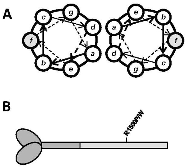
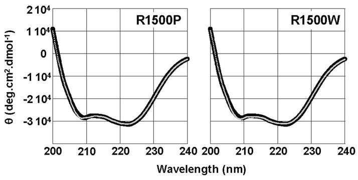
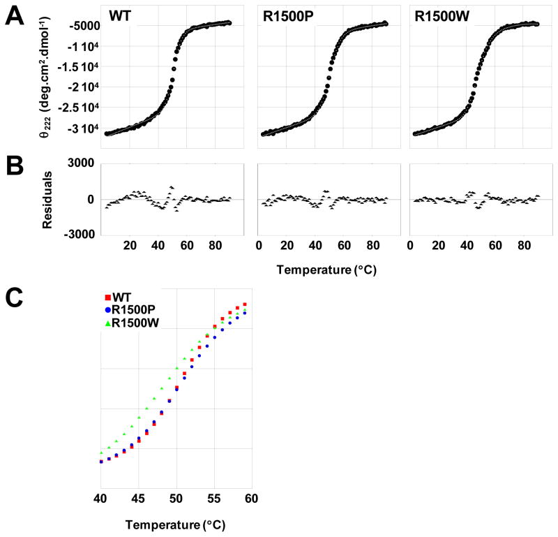
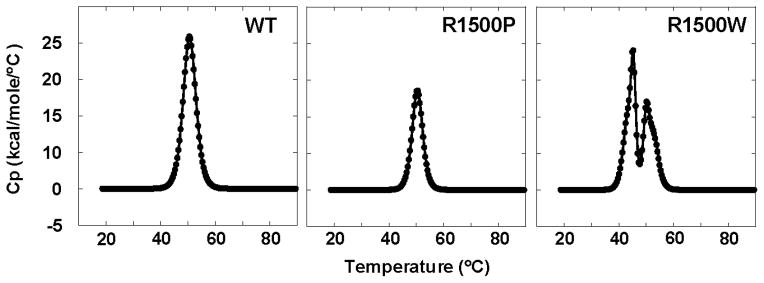
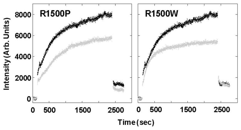
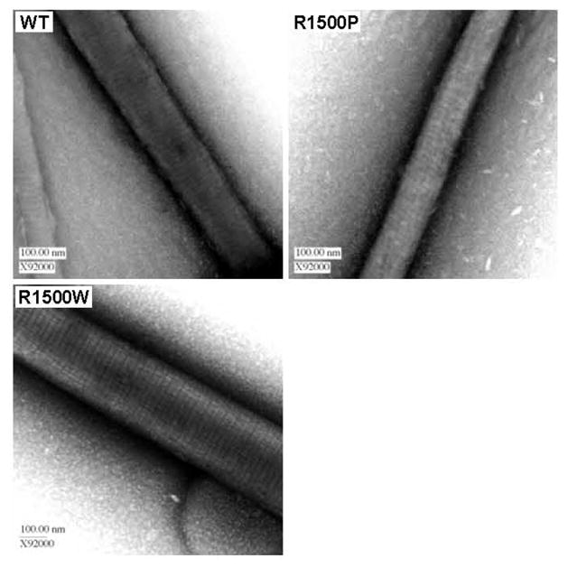
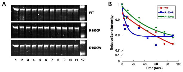
To date, more than 230 disease-causing mutations have been linked to the slow/cardiac muscle myosin gene, beta-MyHC (MYH7). Most of these mutations are located in the globular head region of the protein and result in cardiomyopathies. Recently, however, a number of novel disease-causing mutations have been described in the long, alpha-helical, coiled coil tail region of the beta-MyHC protein. Mutations in this region are of particular interest because they are associated with a multitude of human diseases, including both cardiac and skeletal myopathies. Here, we attempt to dissect the mechanism(s) by which mutations in the rod region of beta-MyHC can cause a variety of diseases by analyzing two mutations at a single amino acid (R1500P and R1500W) which cause two distinct diseases (Laing-type early-onset distal myopathy and dilated cardiomyopathy, respectively). For diseases linked to the R1500 residue, we find that each mutation displays distinct structural, thermodynamic, and functional properties. Both R1500P and R1500W cause a decrease in thermodynamic stability, although the R1500W phenotype is more severe. Both mutations also affect filament assembly, with R1500P causing an additional decrease in filament stability. In addition to furthering our understanding of the mechanism of pathogenesis for each of these diseases, these data also suggest how the variance in molecular phenotype may be associated with the variance in clinical phenotype present with mutations in the beta-MyHC rod.


Figures

Fig 1
Fig 1
Location of R1500P and R1500W mutations in the coiled-coil rod of β-MyHC. (A) Relative amino acid positions within the heptad repeat of a coiled-coil, denoted by a-g. R1500P and R1500W are located in the shaded f position. (B) Diagram of β-MyHC structure. Heavy meromyosin is comprised of the globular head and N-terminal portion of the myosin rod, and is shown in dark gray. LMM is the C-terminal coiled-coil tail region and is shaded light gray, with the relative position of the R1500P and R1500W mutations marked.
Fig 2
Fig 2
R1500P and R1500W mutations do not detectibly alter the secondary structure of LMM. Far-UV CD spectra of WT (black) and mutant (gray) LMM were obtained from 250 nm – 200 nm at 4 °C. WT LMM has nearly identical secondary structure profiles to R1500P and R1500W LMM. All proteins display canonical α-helical spectra with characteristic minima at 208 nm and 222 nm, and the calculated percentage of α-helix is similar for all three proteins.
Fig 3
Fig 3
Thermal denaturation of LMM reveals variation in R1500P and R1500W thermodynamic profiles. (A) The α-helical secondary structure of LMM was monitored by θ222 during thermally denaturation. Measured θ222 data (black) were fit to a theoretical model (gray) to derive thermodynamic parameters. (B) Residuals for the fit of the theoretical model to the measured data are calculated as the difference between the two at each point. Residuals are all around zero, indicating that our model fits well and shows no systematic deviation. (C) Fit θ222 data are plotted over the central segment of denaturation (40 °C to 60 °C) to directly illustrate differences in thermal stability. R1500W LMM (green) melts at a lower temperature and exhibits a wider transition than WT (red) and R1500P (blue) LMM, indicating a decreased cooperativity of melting for R1500W.
Fig 4
Fig 4
DSC thermograms of WT, R1500P, and R1500W LMM. Thermograms show theoretical fits of the data modeled using a two-state transition. Two peaks were deconvoluted for R1500W and thermodynamic parameters calculated separately for each.
Fig 5
Fig 5
R1500P and R1500W LMM display similar defects in self-assembly. Baseline 90° light scattering in no salt buffer was obtained for 120 sec before an equal volume of either WT (Black) or mutant (gray) LMM was added, diluting the sample to a final concentration of 150 mM NaCl and 200 nM protein to initiate self-assembly. Data were collected at a rate of one point/sec, and the reaction was allowed to proceed for 40 min before the addition of 5M salt to return the buffer to 300 mM NaCl and demonstrate the reversibility of the reaction. The intensity of 90° light scattering is plotted in arbitrary units with respect to time.
Fig 6
Fig 6
Ultrastructures of mutant and WT paracrystals are indistinguishable. (A) WT and mutant paracrystals have similar periodicities. WT and mutant LMM were dialyzed overnight into crystallization buffer to induce paracrystal formation. Paracrystals were adsorbed onto carbon mesh grids and electron micrographs were recorded at a magnification of X92000. Paracrystal periodicity measurements were taken over the range of several striations and divided by the number of striations to obtain an average periodicity per paracrystal.
Fig 7
Fig 7
R1500 mutations have differential effects on the proteolytic stability of LMM. (A) Time course for limited proteolytic digestion of LMM. Lane 12 shows molecular weight markers of 125 kDa and 82 kDa. (B) Proteolysis data are plotted as relative band intensity of full length LMM, and fit to an exponential decay curve versus time. The intensity of full length LMM at time zero is defined as 1, and the intensity of the full length LMM band at each time point is plotted with respect to that value.

References

    1. McLachlan AD, Karn J. Periodic charge distributions in the myosin rod amino acid sequence match cross-bridge spacings in muscle. Nature. 1982 Sep 16;299(5880):226–31. - PubMed
    1. Atkinson SJ, Stewart M. Molecular interactions in myosin assembly. Role of the 28-residue charge repeat in the rod. J Mol Biol. 1992 Jul 5;226(1):7–13. - PubMed
    1. Atkinson SJ, Stewart M. Molecular basis of myosin assembly: coiled-coil interactions and the role of charge periodicities. J Cell Sci Suppl. 1991;14:7–10. - PubMed
    1. Buvoli M, Hamady M, Leinwand LA, Knight R. Bioinformatics assessment of beta-myosin mutations reveals myosin’s high sensitivity to mutations. Trends Cardiovasc Med. 2008 May;18(4):141–9. - PMC - PubMed
    1. Armel TZ, Leinwand LA. Mutations in the beta-myosin rod cause myosin storage myopathy via multiple mechanisms. Proc Natl Acad Sci U S A. 2009 Apr 14;106(15):6291–6. - PMC - PubMed

Publication types