

Review
doi: 10.1016/j.visres.2009.10.014.
Epub 2009 Oct 23.
The value of measurement of macular carotenoid pigment optical densities and distributions in age-related macular degeneration and other retinal disorders
Affiliations
- PMID: 19854211
- PMCID: PMC2840187
- DOI: 10.1016/j.visres.2009.10.014
Item in Clipboard
Review
The value of measurement of macular carotenoid pigment optical densities and distributions in age-related macular degeneration and other retinal disorders
Vision Res.
.
Abstract
There is increasing recognition that the optical and antioxidant properties of the xanthophyll carotenoids lutein and zeaxanthin play an important role in maintaining the health and function of the human macula. In this review article, we assess the value of non-invasive quantification of macular pigment levels and distributions to identify individuals potentially at risk for visual disability or catastrophic vision loss from age-related macular degeneration, and we consider the strengths and weaknesses of the diverse measurement methods currently available.
Copyright 2009 Elsevier Ltd. All rights reserved.
Figures
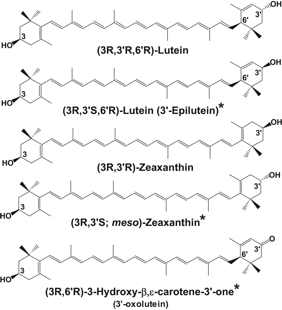
Xanthophyll carotenoids found in the human retina and macula. The asterisks denote metabolites of dietary lutein and zeaxanthin.
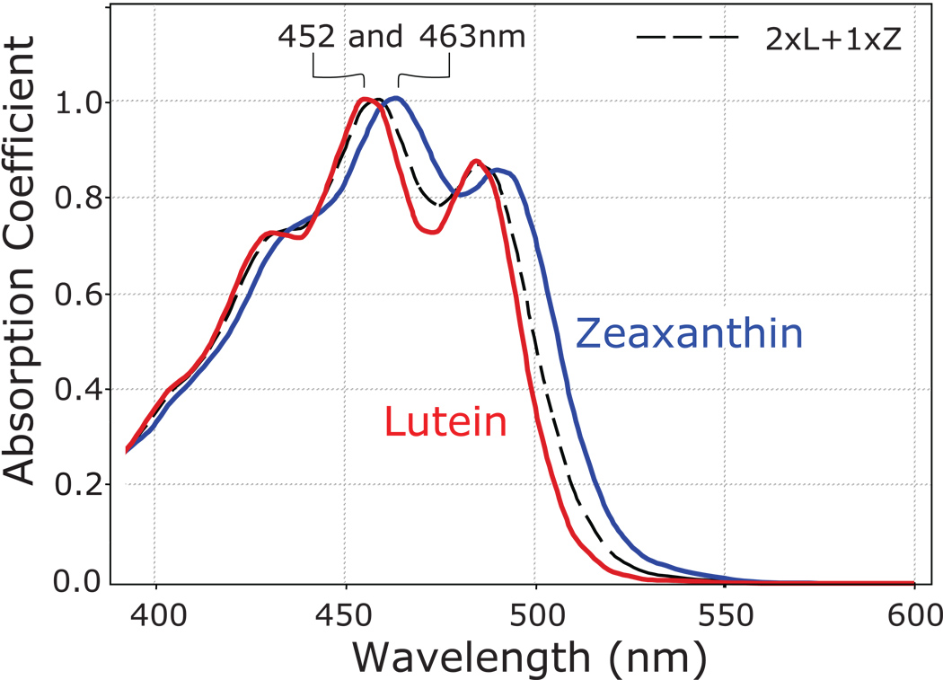
Absorption spectra of lutein (red) and zeaxanthin (blue) in olive oil. A mixture of lutein plus zeaxanthin (dashed black line) closely approximates the absorption spectrum of the macular pigment in the living human eye.
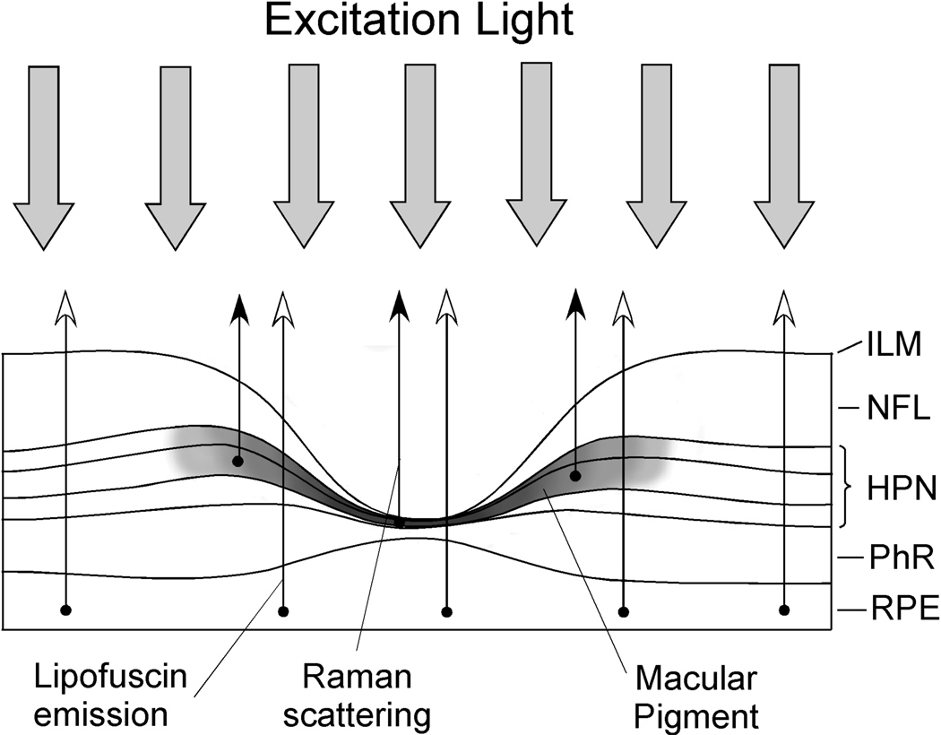
Schematic diagram of the light pathways used in the various methods to measure macular pigment in the living human eye. The dark band corresponds to the location of the macular carotenoids in the fovea. In heterochromatic flicker photometry (HFP), the photoreceptors detect incoming light which is attenuated differentially in the fovea and parafovea depending on the amount of macular pigment encountered. In reflectometry, incoming light is reflected off of various retinal structures of the outer retina, RPE/choroid, and sclera. The double-pass attenuation by the macular pigment is then calculated. In resonance Raman spectroscopy (RRS), incoming light is Raman scattered by the macular carotenoids of the inner retina which is then optically collected and analyzed spectroscopically or displayed in an imaging mode. In autofluorescence imaging (AFI), incoming blue light causes RPE lipofuscin to fluoresce which is subsequently imaged by a scanning laser ophthalmoscope or a CCD camera. The macular pigment of the fovea attenuates the incoming blue light but does not attenuate the longer wavelength fluorescence. This attenuation is imaged and quantified. Abbreviations: inner plexiform layer (ILM); nerve fiber layer (NFL); Henle fiber, plexiform, and nuclear layers (HPN); photoreceptor layer (PhR); retinal pigment epithelium (RPE). (Reprinted from Sharifzadeh, et al., (2006) with permission).
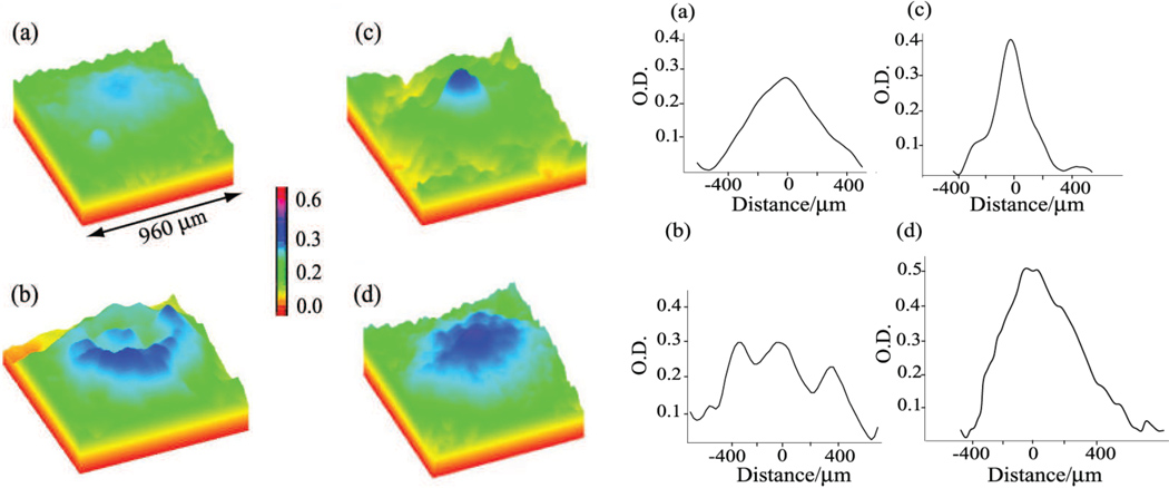
Varied macular pigment distributions measured by single-wavelength autofluorescence imaging. Pseudocolor images of the macular carotenoid pigments from four individuals are on the left showing narrow and broad distributions and ring structures. Line plots along the horizontal axis for the same subjects are shown on the right. (Reprinted from Sharifzadeh, et al., (2006) with permission).
References
-
- Aleman TS, Duncan JL, Bieber ML, de Castro E, Marks DA, Gardner LM, Steinberg JD, Cideciyan AV, Maguire MG, Jacobson SG. Macular pigment and lutein supplementation in retinitis pigmentosa and usher syndrome. Investigative Ophthalmology and Visual Science. 2001;42(8):1873–1881. - PubMed
-
- Alexander KR, Kilbride PE, Fishman GA, Fishman M. Macular pigment and reduced foveal short-wavelength sensitivity in retinitis pigmentosa. Vision Research. 1987;27(7):1077–1083. - PubMed
-
- AREDS Report No. 22. The Relationship of Dietary Carotenoid and Vitamin A, E, and C Intake With Age-Related Macular Degeneration in a Case-Control Study: AREDS Report No. 22. Archives of Ophthalmology. 2007;125(9):1225–1232. - PubMed
Publication types
MeSH terms
Substances
Grants and funding
LinkOut - more resources
Full Text Sources
Other Literature Sources
Medical
