. 2010 Feb;31(4):779-91.
doi: 10.1016/j.biomaterials.2009.09.082. Epub 2009 Oct 24.

Long-term biostability of self-assembling protein polymers in the absence of covalent crosslinking

Affiliations

Long-term biostability of self-assembling protein polymers in the absence of covalent crosslinking

Rory E Sallach et al. Biomaterials. 2010 Feb.

Abstract

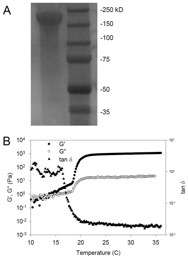
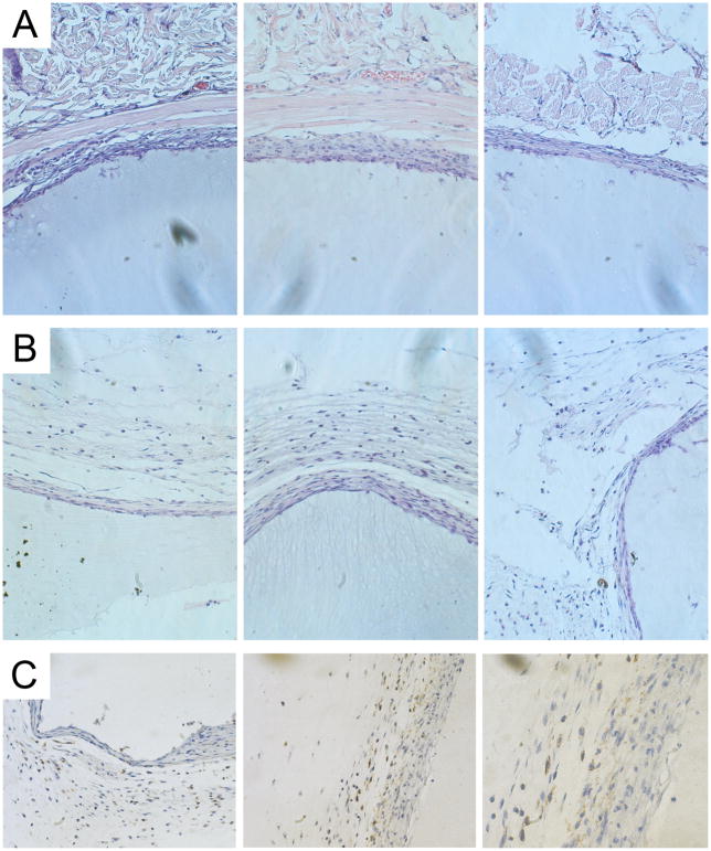
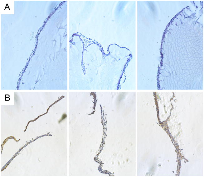
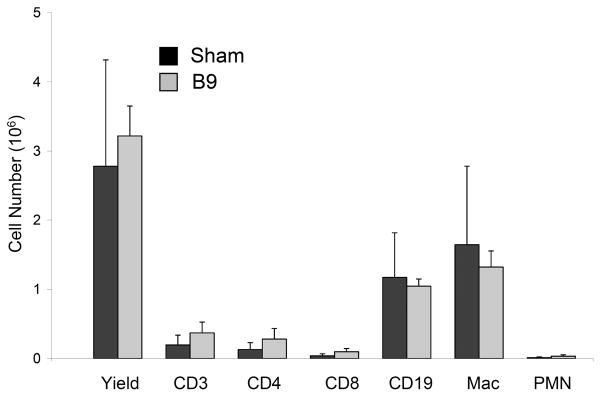
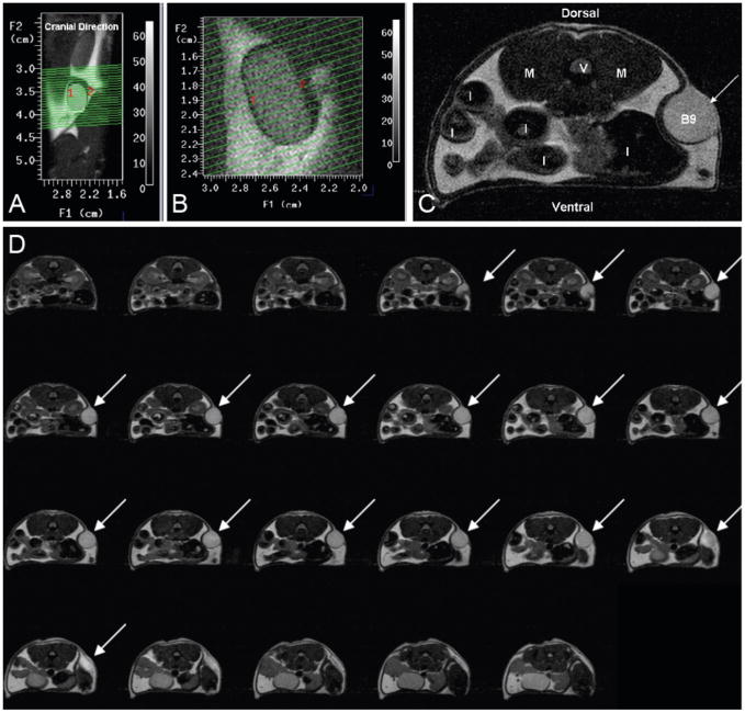
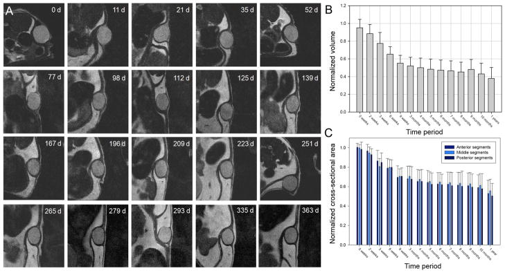
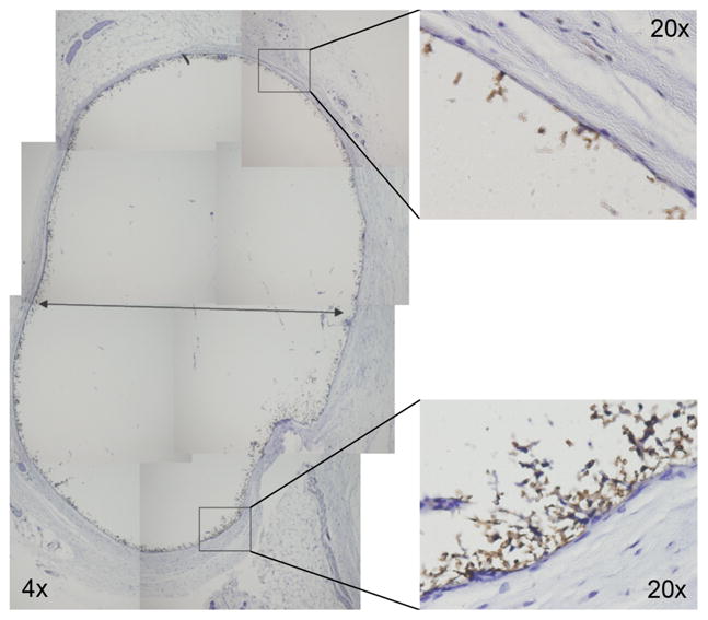
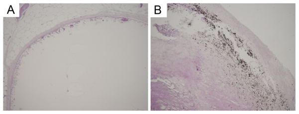
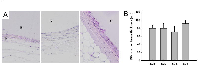
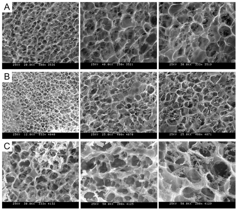
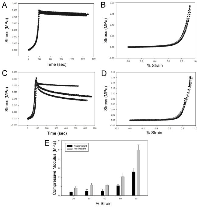
Unless chemically crosslinked, matrix proteins, such as collagen or silk, display a limited lifetime in vivo with significant degradation observed over a period of weeks. Likewise, amphiphilic peptides, lipopeptides, or glycolipids that self-assemble through hydrophobic interactions to form thin films, fiber networks, or vesicles do not demonstrate in vivo biostability beyond a few days. We report herein that a self-assembling, recombinant elastin-mimetic triblock copolymer elicited minimal inflammatory response and displayed robust in vivo stability for periods exceeding 1 year, in the absence of either chemical or ionic crosslinking. Specifically, neither a significant inflammatory response nor calcification was observed upon implantation of test materials into the peritoneal cavity or subcutaneous space of a mouse model. Moreover, serial quantitative magnetic resonance imaging, evaluation of pre- and post-explant ultrastructure by cryo-high resolution scanning electron microscopy, and an examination of implant mechanical responses revealed substantial preservation of form, material architecture, and biomechanical properties, providing convincing evidence of a non-chemically or ionically crosslinked protein polymer system that exhibits long-term stability in vivo.


Figures

Figure 1
Figure 1. Structural characterization of a triblock elastin-mimetic protein polymer
(A) Sodium dodecyl sulfate-polyacrylamide gel electrophoresis (SDS-PAGE) analysis revealed a single protein band at 170 KDa corresponding to B9. A total of 10 μg of the elastin-mimetic polypeptide was run on 7.5% gel and negatively stained with Copper stain (Bio-Rad). Molecular weight markers were Precision Plus Protein Kaleidoscope (Bio-Rad). (B) Rheological behavior of 10-wt % B9 in water. Dynamic shear storage (G′) and loss modulus (G″) are plotted as a function of temperature (γ2%, ω 1Hz). The gelation temperature was determined by heating samples from 4°C to 37°C at a rate of 1°C per minute. Experiments were repeated on three samples and representative data presented.
Figure 2
Figure 2. Short-term host-implant responses after subcutaneous injection of protein polymer in a mouse model
A 10 wt% solution of B9 was injected subcutaneously, which gelled instantly. Samples were retrieved at 3 weeks and hematoxylin and eosin staining performed on formalin fixed, paraffin embedded implants. Histological analysis of the host response to the subcutaneous protein polymer implant was conducted at the implant-muscle (A) and implant-dermal (B) tissue interfaces. (C) F4/80 staining demonstrates the presence of macrophages along the periphery of the fibrous capsule but no infiltration into the implant. All images were obtained at 20x magnification.
Figure 3
Figure 3. Short-term host-implant responses after peritoneal injection of protein polymer in a mouse model
(A) Histological analysis of hematoxylin and eosin stained formalin fixed, paraffin embedded peritoneal implants retrieved 1 week after implantation. (B) F4/80 staining of implants demonstrates the presence of macrophages along the periphery of the implant. All images were obtained at 20x magnification.
Figure 4
Figure 4. FACS analysis of peritoneal cell response to protein polymer implant 1 week after implantation
Cells from the peritoneal lavage were immunostained for flow cytometry with FITC-conjugated hamster anti-mouse CD3 for total T cells, FITC-conjugated rat monoclonal anti-mouse CD4 for helper T cells, FITC-conjugated rat monoclonal anti-mouse CD8 for cytotoxic T cells, FITC-conjugated rat monoclonal anti-mouse CD19 for B cells, PE-conjugated rat monoclonal anti-mouse CD11b for macrophage, and FITC-conjugated rat monoclonal anti-mouse Gr-1 for neutrophils. There was no observable difference in cellular response between mice receiving the implant or those that underwent sham surgery (n=5, p < 0.05).
Figure 5
Figure 5. MR imaging of a subcutaneously implanted protein polymer cylindrical implant
(A) A coronal view through the mouse, oriented vertically, shows the location of the implant. The green lines depict 23 transverse-oblique slices through the implant. (B) MR scan slices through implant. (C) Transverse MR image of the subcutaneous B9 implant. (V = verterbra, M = Psoas Major muscle, I = Intestine cross-sections, B9 = cross-section of B9 implant). (D) Sequential series of transverse MR images through a subcutaneously placed B9 implant. Implant areas were assessed from individual images and summed to assess volume of the implant. Slice thickness is 500 μm.
Figure 6
Figure 6. Serial MR imaging of subcutaneous protein polymer implants over a 12 month period
(A) Representative serial images of B9 implants over a 1-year period. Normalized implant volumes (B) and implant cross-sectional areas (C) were determined over a 1-year implant period (n = 7, mean ± SD).
Figure 7
Figure 7. Histological analysis of protein polymer implants retrieved 1 year after implantation
F4/80 staining of formalin fixed, paraffin embedded B9 implants demonstrate the presence of macrophages along the periphery of the fibrous capsule but no infiltration into the B9 implant. Whole implant image was obtained at 4x magnification, while high power views were obtained at 20x magnification.
Figure 8
Figure 8. Histological analysis of protein polymer implants retrieved 1 year after implantation
Von Kossa staining does not reveal the presence of calcium in the B9 implant (left), but demonstrates calcium deposits in a human carotid atherosclerotic plaque (right), used as a positive control. Images were obtained at 4x magnification.
Figure 9
Figure 9. Analysis of fibrous capsule response to implants retrieved 1 year after implantation
(A) Histological examination of protein polymer B9 implanted in the dorsal subcutaneous space of C57BL/6 mice for 1 year. A fibrous capsule (F) was noted surrounding the protein polymer (G). Samples were retrieved with capsule intact and hematoxylin and eosin staining performed on formalin fixed, paraffin embedded implants. (B) Capsule thickness was determined from measurements obtained from images obtained at 20x magnification from at least six distinct regions on each surrounding capsule in which each region is represented by a minimum of 10 high power fields. The capsule thickness is presented for four separate implants designated SC1 through SC4. The overall mean thickness ± standard deviation for all samples was 80 ± 8 μm.
Figure 10
Figure 10. Cryo-HRSEM micrographs of protein polymer implants
(A) Images of hydrated 10-wt% B9 implants prior to implantation. After a 1-year implant period, implants were retrieved and the capsule removed. Cryo-HRSEM was performed to visualized the internal structure (B) of the implant after fracturing specimens with a pre-chilled blade and of the surface (C) of the implants. Magnification ranges from 25,000 to 50,000x.
Figure 11
Figure 11. Biomechanical analysis of compression stress-strain responses of implants retrieved 1 year after implantation
Mechanical responses of protein polymer samples were characterized by compression testing prior to implantation (A, B) and at the time of specimen removal, one year after initial implantation (C, D, E). Cylindrical specimens were fabricated and cut to a thickness of 2.35 mm for mechanical testing in a Bose ELF system in a hydration chamber containing PBS at 37°C. Samples were preconditioned by cyclic compression ranging between 1% and 20% strain for 10 cycles. Compression stress-relaxation testing was conducted by compressing the sample to a strain of 75% at 0.025 mm/sec followed by monitoring change in compression stress over time (A, C). Compression stress-strain testing was performed at 0.025 mm/sec (B, D). Cotangent moduli (E) were calculated from the compression stress-strain plots at 10% strain intervals between 20% and 60% strain.

References

    1. Wright ER, Conticello VP. Self-assembly of block copolymers derived from elastin-mimetic polypeptide sequences. Adv Drug Deliv Rev. 2002;54:1057–1073. - PubMed
    1. Wright ER, McMillan RA, Cooper A, Apkarian RP, Conticello VP. Thermoplastic elastomer hydrogels via self-assembly of an elastin-mimetic triblock polypeptide. Advanced Functional Materials. 2002;12(2):1–6.
    1. Nagapudi K, Brinkman WT, Thomas BS, Wright ER, Conticello VP, Chaikof EL. Protein-based thermoplastic elastomers. Macromolecules. 2005;38:345–354.
    1. Wu X, Sallach R, Haller CA, Caves JA, Nagapudi K, Conticello VP, et al. Alterations in physical cross-linking modulate mechanical properties of two-phase protein polymer networks. Biomacromolecules. 2005;6(6):3037–3044. - PubMed
    1. Wu X, Sallach RE, Caves JM, Conticello VP, Chaikof EL. Deformation responses of a physically cross-linked high molecular weight elastin-like protein polymer. Biomacromolecules. 2008;9:1787–1794. - PMC - PubMed

Publication types