. 2009 Dec;150(12):5626-32.
doi: 10.1210/en.2009-0881. Epub 2009 Oct 23.

Transgenic mice expressing green fluorescent protein under the control of the corticotropin-releasing hormone promoter

Affiliations

Transgenic mice expressing green fluorescent protein under the control of the corticotropin-releasing hormone promoter

Tamar Alon et al. Endocrinology. 2009 Dec.

Abstract

CRH is widely expressed in the brain and is of broad functional relevance to a number of physiological processes, including stress response, parturition, immune response, and ingestive behavior. To delineate further the organization of the central CRH network, we generated mice expressing green fluorescent protein (GFP) under the control of the CRH promoter, using bacterial artificial chromosome technology. Here we validate CRH-GFP transgene expression within specific brain regions and confirm the distribution of central GFP-producing cells to faithfully recapitulate that of CRH-expressing cells. Furthermore, we confirm the functional integrity of a population of GFP-producing cells by demonstrating their opposite responsiveness to nutritional status. We anticipate that this transgenic model will lend itself as a highly tractable tool for the investigation of CRH expression and function in discrete brain regions.


Figures
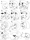
Figure 1
Figure 1
Schematic representation of GFP-IR in CRH-GFP mouse brain. A series of representative rostrocaudally aligned schematic transverse sections (A–L) depicting the location of the neurons labeled for GFP-IR (black circles) in the CRH-GFP mouse brain. Each black circle represents a single GFP-IR neuron. 3V, Third ventricle; 4V, fourth ventricle; 7n, facial nerve; II/III, cerebral cortex, layer 2 and 3; V/VI, cerebral cortex, layer 5 and 6; ac, anterior commissure; AcbSh, shell part of accumbens nucleus; Aq, aqueduct; AP, area postrema; Bar, Barrington’s nucleus; BMA, anterior part of basomedial amygloid nucleus; BSTLD, lateral division of bed nucleus of stria terminalis, dorsal part; BSTLV, lateral division of bed nucleus of stria terminalis, ventral part; CA, hippocampus; CC, central canal; CeA, central nucleus of the amygdala; Cg, cingulate cortex; cp, cerebral peduncle, basal part; CPu, caudate putamen; D3V, dorsal third ventricle; DG, dentate gyrus; DMD, dorsal part of the dorsomedial nucleus of the hypothalamus; DpMe, deep mesencephalic nucleus; f, fornix; fmi, forceps minor of the corpus callosum; fr, fasciculus retroflexus; GiA, gigantocellular reticular nucleus, α ic, internal capsule; IC, inferior colliculus; IL, infralimbic cortex; IO, inferior olive; IPAC, interstitial nucleus of the posterior limb of the anterior commissure; LDTg, laterodorsal tegmental nucleus; LRt, lateral reticular nucleus; LPB, lateral parabrachial nucleus; LS, lateral septal nucleus; LV, lateral ventricle; MeA, medial amygdaloid nucleus; MGM, medial geniculate nucleus; ml, medial lemniscus; MM, medial mammillary nucleus, medial part; Mo, motor cortex; MPO, medial preoptic area; MS, medial septal nucleus; mt, mammillothalamic tract; MVe, medial vestibular nucleus; RMg, raphe magnus nucleus; RgTg, reticulotegmental nucleus of the pons; PAG, pariaqueductal gray; PaLM, lateral magnocellular pat of the paraventricular nucleus of hypothalamus; Pir, piriform cortex; PnO, pontine reticular nucleus, oral part; PPTg, pedunculopotine tegmental nucleus; RMg, raphe magnus nucleus; SI, substantia innominata; SNc, substantia nigra, compact part; SNr, substatia nigra, reticular part; sol, nucleus of solitary tract; SS, somatosensory cortex; st, stria terminalis; Tu, olfactory tubercle; VP, ventral pallidum; XSCP, decussation of the superior cerebellar peduncle; ZI, zona incerta. Scale bar (L), 2 mm.
Figure 2
Figure 2
Colocalization of GFP-IR and CRH-IR (A–J“) and GFP-IR and 35S-labeled CRH (K–T) in CRH-GFP mouse brain. A–E, Merged photomicrographs of representative regions (BSTLD, PVH, LHA, A11, and Barrington’s nucleus, respectively) expressing GFP-IR (green fluorescence) and CRH-IR (red fluorescence). F–J, Higher-power magnification of boxed area in A–E, respectively, and indicate neurons expressing GFP-IR. F’–J’, Neurons express CRH-IR. F”–J“, Merged images of GFP-IR and CRH-IR. Arrows indicate colocalization of GFP-IR and CRH-IR. Scale bar (A), 300 μm applies to A; scale bar (E), 300 μm applies to B–E; scale bar (J”), 25 μm, applies to all other micrographs. K–O, Photomicrographs of representative regions (BST, PVH, LHA, A11, and Barrington’s nucleus, respectively) expressing GFP-IR (brown stain) and 35S-labeled CRH (cluster of black grains). P–T, Higher-power magnification of boxed area (K–O), respectively, and illustrate neurons coexpressing GFP-IR and 35S-labeled CRH. Scale bar (O), 300 μm applies to K–O; scale bar (T), 25 μm, applies to P–T. For abbreviation definition, see legend to Fig. 1.
Figure 3
Figure 3
Refeeding substantially activates GFP neurons in CRH-GFP mice. A, Merged photomicrographs of FOS-IR (green fluorescence), used as a marker of neuronal activation, and GFP-IR (red fluorescence) in the PVH of CRH-GFP mice after 18 h food deprivation proceeded by a 4-h bout of refeeding. B, Higher-power magnification of PVH FOS-IR neurons. B’, The same section as in B but illustrating GFP-IR. B“, The merged photomicrograph of B and B’ illustrating coexpression of FOS-IR and GFP-IR in a representative sample of PVH neurons. Arrows indicate colocalization. Scale bar (A), 100 μm applies to A; scale bar (B”), 25 μm, applies to B–B“. For abbreviation definition, see legend to Fig. 1.

References

    1. Bamberger CM, Bamberger AM 2000 The peripheral CRH/urocortin system. Ann NY Acad Sci 917:290–296 - PubMed
    1. Keegan CE, Herman JP, Karolyi IJ, O'Shea KS, Camper SA, Seasholtz AF 1994 Differential expression of corticotropin-releasing hormone in developing mouse embryos and adult brain. Endocrinology 134:2547–2555 - PubMed
    1. Merchenthaler I, Vigh S, Petrusz P, Schally AV 1982 Immunocytochemical localization of corticotropin-releasing factor (CRF) in the rat brain. Am J Anat 165:385–396 - PubMed
    1. Potter E, Sutton S, Donaldson C, Chen R, Perrin M, Lewis K, Sawchenko PE, Vale W 1994 Distribution of corticotropin-releasing factor receptor mRNA expression in the rat brain and pituitary. Proc Natl Acad Sci USA 91:8777–8781 - PMC - PubMed
    1. Sawchenko PE, Swanson LW, Vale WW 1984 Corticotropin-releasing factor: coexpression within distinct subsets of oxytocin-, vasopressin-, and neurotensin-immunoreactive neurons in the hypothalamus of the male rat. J Neurosci 4:1118–1129 - PMC - PubMed

Publication types

MeSH terms

Substances