. 2010 Jan;192(1):94-103.
doi: 10.1128/JB.00848-09.

Cytoplasmic histidine kinase (HP0244)-regulated assembly of urease with UreI, a channel for urea and its metabolites, CO2, NH3, and NH4(+), is necessary for acid survival of Helicobacter pylori

Affiliations

Cytoplasmic histidine kinase (HP0244)-regulated assembly of urease with UreI, a channel for urea and its metabolites, CO2, NH3, and NH4(+), is necessary for acid survival of Helicobacter pylori

David R Scott et al. J Bacteriol. 2010 Jan.

Abstract

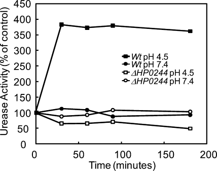
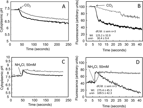
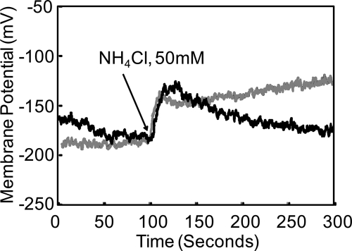
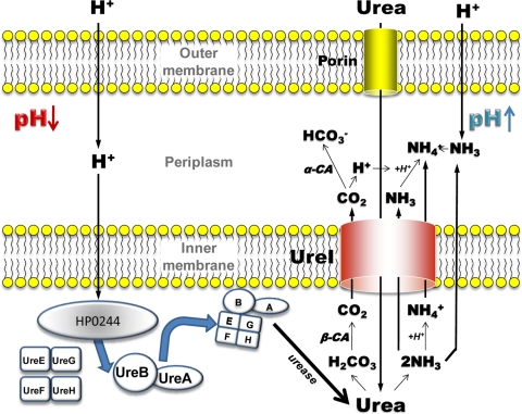
Helicobacter pylori colonizes the normal human stomach by maintaining both periplasmic and cytoplasmic pH close to neutral in the presence of gastric acidity. Urease activity, urea flux through the pH-gated urea channel, UreI, and periplasmic alpha-carbonic anhydrase are essential for colonization. Exposure to pH 4.5 for up to 180 min activates total bacterial urease threefold. Within 30 min at pH 4.5, the urease structural subunits, UreA and UreB, and the Ni(2+) insertion protein, UreE, are recruited to UreI at the inner membrane. Formation of this complex and urease activation depend on expression of the cytoplasmic sensor histidine kinase, HP0244. Its deletion abolishes urease activation and assembly, impairs cytoplasmic and periplasmic pH homeostasis, and depolarizes the cells, with an approximately 7-log loss of survival at pH 2.5, even in 10 mM urea. Associated with this assembly, UreI is able to transport NH(3), NH(4)(+), and CO(2), as shown by changes in cytoplasmic pH following exposure to NH(4)Cl or CO(2). To be able to colonize cells in the presence of the highly variable pH of the stomach, the organism expresses two pH-sensor histidine kinases, one, HP0165, responding to a moderate fall in periplasmic pH and the other, HP0244, responding to cytoplasmic acidification at a more acidic medium pH. Assembly of a pH-regulatory complex of active urease with UreI provides an advantage for periplasmic buffering.


Figures

FIG. 1.
FIG. 1.
Urease activity of intact H. pylori incubated in acid. H. pylori wild-type (Wt) and HP0244 deletion mutant strains were incubated in BHI, pH 4.5 or 7.4, for 30 to 180 min, and total urease activity was measured in the presence of a nonionic detergent to bypass UreI. In the wild-type strain, there was a >3-fold increase in urease activity at pH 4.5 compared to that at pH 7.4 after 30 min, and it remained elevated for 3 h. The absence of HP0244 had no effect on urease activity at pH 7.4 but reduced urease activity about 50% with incubation at pH 4.5 within 30 min and further reduced the activity at pH 4.5 after 180 min.
FIG. 2.
FIG. 2.
Western blot analysis and urease activity of purified membrane fractions from wild-type and HP0244 deletion strains of H. pylori. Representative Western blots are shown for UreA, UreB, UreE, and UreI from wild-type (A) and HP0244 deletion (B) strains that were exposed to pH 7.4 and pH 4.5. The intensities of the protein bands were measured and converted to expression ratios of pH 4.5 to pH 7.4. (C) The amounts of UreA, UreB, and UreE but not UreI protein increased after incubation at pH 4.5 for 30 min in wild-type H. pylori, whereas the absence of HP0244 abolished the recruitment of UreA and UreB, but not UreE, to the membrane. (D) Urease activities from both membrane and soluble fractions incubated at pH 4.5 and pH 7.4 from wild-type and HP0244 deletion strains were measured. The urease activity of the membrane fraction increased ∼2-fold after incubation at pH 4.5 for 30 min in the wild-type strain (black bars) but not in the HP0244 deletion strain (gray bars). Urease activity is expressed as a percentage of that in the pH 7.4 control.
FIG. 3.
FIG. 3.
Deletion of HP0244 abolishes periplasmic buffering in H. pylori. Wild-type (A) and HP0244 deletion (B) strains were incubated at pH 5.0 in the presence of BCECF free acid to monitor periplasmic pH changes. Representative images show that the addition of 5 mM urea resulted in increased peripheral fluorescence due to increased periplasmic pH in the wild type but not in the HP0244 deletion mutant. Arrows indicate periplasmic fluorescence.
FIG. 4.
FIG. 4.
UreI is permeable to CO2, NH3, and NH4+. (A) In the H. pylori wild-type strain (black line), CO2 addition rapidly decreased pHin, followed by a slower decrease in pHin. Deletion of ureI (gray line) greatly reduced the initial rapid fall in pHin but had no effect on the slow phase of pHin acidification. The initial rapid acidification phase is due to CO2 entry through UreI, and the secondary, slow phase of cytoplasmic acidification is due to permeation of CO2 through the lipid bilayer, reflecting continued CO2 addition to the medium. (B) Single-wavelength (502 nm) excitation of BCECF was used for rapid measurement (data capture every 50 ms) of the rate of change of fluorescence. The rate of cytoplasmic fluorescence decrease (alkalization) following CO2 addition was significantly higher in the wild-type organism (black line) than in the ureI deletion mutant (gray line), showing that UreI is able to accelerate CO2 movement across the inner membrane of H. pylori (dF/dt ± standard error of the mean [SEM]) (n = 3; P ≤ 0.05). (C) NH4Cl addition rapidly increases pHin due to NH3 entry, followed by fall in pHin, in the H. pylori wild-type strain (black line). Deletion of ureI (gray line) abolished the secondary fall in pHin. (D) The rate of cytoplasmic fluorescence increase (alkalization) following NH4Cl addition was significantly higher in the wild-type organism (black line) than in the ureI deletion mutant (gray line), showing that UreI is able to accelerate NH4+ movement across the inner membrane of H. pylori (dF/dt ± SEM) (n = 3; P ≤ 0.05). Therefore, the secondary fall in pHin is due to NH4+ entry through UreI, with generation of H+ in the bacterial cytoplasm (dF/dt ± SEM) (n = 3; P ≤ 0.05).
FIG. 5.
FIG. 5.
Changes of membrane potential following addition of NH4Cl. In the wild-type and mutant organisms, there was an immediate fall in potential due to alkalization of the interior, as shown in Fig. 4C, with an increase in ΔpH. In the wild-type strain, repolarization is due to a fall in pHin that is absent in the HP0244 deletion mutant, arguing for NH4+ permeation of UreI.
FIG. 6.
FIG. 6.
Model of the mechanism of action of UreI and HP0244. UreI transports urea at acidic periplasmic pH (pH < 6.2) and increasing urease activity, forming H2CO3 and 2 NH3. Carbonic acid is converted to CO2 in the cytoplasm by β-carbonic anhydrase and enters the periplasm, where it is converted by α-carbonic anhydrase to HCO3+, enabling maintenance of periplasmic pH at ∼6.1. NH3 exits via the bilayer and UreI, and NH4+ is formed from the H+ generated by α-carbonic anhydrase and from protons entering from the medium. The NH4+ generated in the cytoplasm exits via UreI, or perhaps via NH3 + H+ exit. Acidification of the cytoplasm activates HP0244, which in turn allows assembly of the apoenzyme UreA/UreB with the nickel insertion pairs, UreE/UreG and UreF/UreH, activating urease at the membrane, providing local generation of carbonic acid and ammonia.

Similar articles

Cited by

References

    1. Alm, R. A., L. S. Ling, D. T. Moir, B. L. King, E. D. Brown, P. C. Doig, D. R. Smith, B. Noonan, B. C. Guild, B. L. deJonge, G. Carmel, P. J. Tummino, A. Caruso, M. Uria-Nickelsen, D. M. Mills, C. Ives, R. Gibson, D. Merberg, S. D. Mills, Q. Jiang, D. E. Taylor, G. F. Vovis, and T. J. Trust. 1999. Genomic-sequence comparison of two unrelated isolates of the human gastric pathogen Helicobacter pylori. Nature 397:176-180. - PubMed
    1. Athmann, C., N. Zeng, T. Kang, E. A. Marcus, D. R. Scott, M. Rektorschek, A. Buhmann, K. Melchers, and G. Sachs. 2000. Local pH elevation mediated by the intrabacterial urease of Helicobacter pylori cocultured with gastric cells. J. Clin. Invest. 106:339-347. - PMC - PubMed
    1. Beier, D., and R. Frank. 2000. Molecular characterization of two-component systems of Helicobacter pylori. J. Bacteriol. 182:2068-2076. - PMC - PubMed
    1. Bury-Mone, S., G. L. Mendz, G. E. Ball, M. Thibonnier, K. Stingl, C. Ecobichon, P. Ave, M. Huerre, A. Labigne, J. M. Thiberge, and H. De Reuse. 2008. Roles of alpha and beta carbonic anhydrases of Helicobacter pylori in the urease-dependent response to acidity and in colonization of the murine gastric mucosa. Infect. Immun. 76:497-509. - PMC - PubMed
    1. Clyne, M., A. Labigne, and B. Drumm. 1995. Helicobacter pylori requires an acidic environment to survive in the presence of urea. Infect. Immun. 63:1669-1673. - PMC - PubMed

Publication types

MeSH terms

LinkOut - more resources