. 2010 Jan;192(1):217-24.
doi: 10.1128/JB.01124-09.

Metabolic control of virulence genes in Brucella abortus: HutC coordinates virB expression and the histidine utilization pathway by direct binding to both promoters

Affiliations

Metabolic control of virulence genes in Brucella abortus: HutC coordinates virB expression and the histidine utilization pathway by direct binding to both promoters

Rodrigo Sieira et al. J Bacteriol. 2010 Jan.

Abstract

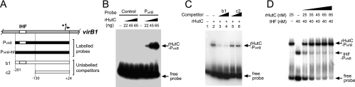
Type IV secretion systems (T4SS) are multicomponent machineries involved in the translocation of effector molecules across the bacterial cell envelope. The virB operon of Brucella abortus codes for a T4SS that is essential for virulence and intracellular multiplication of the bacterium in the host. Previous studies showed that the virB operon of B. abortus is tightly regulated within the host cells. In order to identify factors implicated in the control of virB expression, we searched for proteins of Brucella that directly bind to the virB promoter (P(virB)). Using different procedures, we isolated a 27-kDa protein that binds specifically to P(virB). This protein was identified as HutC, the transcriptional repressor of the histidine utilization (hut) genes. Analyses of virB and hut promoter activity revealed that HutC exerts two different roles: it acts as a coactivator of transcription of the virB operon, whereas it represses the hut genes. Such activities were observed both intracellularly and in bacteria incubated under conditions that resemble the intracellular environment. Electrophoresis mobility shift assays (EMSA) and DNase I footprinting experiments revealed the structure, affinity, and localization of the HutC-binding sites and supported the regulatory role of HutC in both hut and virB promoters. Taken together, these results indicate that Brucella coopted the function of HutC to coordinate the Hut pathway with transcriptional regulation of the virB genes, probably as a way to sense its own metabolic state and develop adaptive responses to overcome intracellular host defenses.


Figures

FIG. 1.
FIG. 1.
Analysis of binding of rHutC to PvirB. (A) Schematic representation of genomic sequences corresponding to PvirB (gray bar) and the first gene of the virB operon (virB1) (striped bar), radioactive labeled probes used for EMSA (black bars), and unlabeled competitors (white bars). White box, IHF-binding site. (B) Specificity of binding of rHutC to PvirB. EMSA performed with rHutC and a control probe or probe PvirB. (C) Competition assay. EMSA performed with probe PvirB, unlabeled competitors, and rHutC. The mass of unlabeled competitors added to each reaction mixture was as follows: lanes 1 and 2, no competitor; lanes 3 and 5, 150 ng; lanes 4 and 6, 300 ng. Mass of rHutC: lane 1, no rHutC; lanes 2 to 6, 33 ng. (D) Coincubation of rHutC and rIHF with PvirB. EMSA performed with probe PvirB, rIHF, and/or rHutC at the indicated concentrations.
FIG. 2.
FIG. 2.
Analysis of binding affinity of HutC to PvirB or Phut. (A) EMSA performed with probe PvirB and increasing concentrations of rHutC. (B) Determination of the apparent dissociation constant of rHutC for the binding to PvirB. The intensity of the bands obtained from two independent experiments performed as shown for panel A was measured. The apparent dissociation constant (Kd) was determined graphically from the plot of fractions of free and protein-bound probes. (C) EMSA performed with probe Phut and increasing concentrations of rHutC. (D) Determination of the apparent dissociation constant of rHutC for binding to Phut. The intensity of the bands obtained from two independent experiments performed as shown for panel C was measured, and the apparent dissociation constant (Kd) was determined as described for panel B.
FIG. 3.
FIG. 3.
Effect of UCA on DNA-binding activity of HutC. (A) EMSA performed with rIHF or rHutC incubated with probe PvirB and increasing concentrations of UCA as indicated. Concentration of rIHF, 70 nM; concentration of rHutC, 25 nM. (B) EMSA performed with rHutC incubated with probe Phut and increasing concentrations of UCA as indicated. Concentration of rHutC, 0.55 nM.
FIG. 4.
FIG. 4.
Identification of the HutC-binding sites. (A) DNase I footprinting experiment carried out with probe Phut and increasing concentrations of HutC as indicated. Lanes A and G show results from DNA sequence reactions performed by the Sanger method. The HutC-protected region is indicated by an open rectangle. Arrowheads indicate DNase I-hypersensitized sites. (B) DNase I footprinting experiment carried out with probe PvirB and increasing concentrations of HutC as indicated. The HutC-protected region and hypersensitized sites are indicated as described for panel A. (C) Schematic representation of the HutC-protected sequences in Phut. The protected region and DNase I-hypersensitized sites are indicated as described for panel A. Nucleotides that match the previously described HutC-binding sites of Klebsiella or Pseudomonas are highlighted in gray. (D) Schematic representation of the HutC-protected sequences in PvirB. The protected region and DNase I-hypersensitized sites are indicated as described for panel A. IHF-binding site sequences are indicated with bold, italic letters. Nucleotides that match the previously described HutC-binding sites are indicated as described for panel C. (E) Comparison of the 14-bp sequences located at the center of the HutC-protected regions of B. abortus Phut and PvirB, the HutC-binding sites of the hut operators of Klebsiella and Pseudomonas, and sequences located within the hut promoter regions of different closely related alphaproteobacterial species. The dyad symmetry of the sequences is indicated by arrows.
FIG. 5.
FIG. 5.
Role of HutC in control of activity of the virB and hut promoters. (A and B) Schematic representation of the PvirB-lacZ (A) and Phut-lacZ (B) transcriptional fusion constructs. Orientation relative to the hutFC and hutIHUG operons is indicated. (C) Intracellular β-Gal activity of strain B. abortus PvirB-lacZ (wt), B. abortus ΔhutC PvirB-lacZhutC), or B. abortus ΔhutC-KI PvirB-lacZ (KI). Cultures of J774 macrophages were infected with strains harboring lacZ transcriptional fusions. At 5 h p.i., cells were disrupted and β-galactosidase activity of intracellular bacteria was determined. (D) Intracellular β-Gal activity of strain B. abortus Phut-lacZ (wt), B. abortus ΔhutC Phut-lacZhutC), or B. abortus ΔhutC-KI Phut-lacZ (KI). Intracellular β-galactosidase activity was determined as described for panel C. (E) β-Gal activity of strain B. abortus PvirB-lacZ (wt), B. abortus ΔhutC PvirB-lacZhutC), or B. abortus ΔhutC-KI PvirB-lacZ (KI) cultured in MM1 and 5 mM UCA at pH 4.5. The promoter activity of cultured bacteria was determined as follows. Strains were grown in rich medium (TSB) until the exponential phase (OD600 of 0.5 to 1). Subsequently, bacteria were harvested and suspended in MM1 and 5 mM UCA, and β-galactosidase activity was determined after 4 h of cultivation. (F) β-Gal activity of strain B. abortus Phut-lacZ (wt), B. abortus ΔhutC Phut-lacZhutC), or B. abortus ΔhutC-KI Phut-lacZ (KI) cultured in MM1 and 5 mM UCA at pH 4.5. The promoter activity of cultured bacteria was determined as described for panel C. Values are means ± standard deviations of duplicate wells from a representative of two experiments. *, P < 0.05; **, P < 0.01 (compared to the wild-type strain).
FIG. 6.
FIG. 6.
(A) Intracellular replication of B. abortus 2308 and the deletion mutant B. abortus ΔhutC in J774 macrophages. Macrophages (1 × 10⁵ per well) were inoculated with 5 × 10⁶ CFU of bacteria. After 1 h of incubation at 37°C, cells were washed with PBS, and gentamicin and streptomycin were added. CFU were determined at the indicated times. Open circles, B. abortus 2308; filled circles, B. abortus ΔhutC. (B) Persistence of B. abortus 2308, the deletion mutant B. abortus ΔhutC, and the knock-in control strain B. abortus ΔhutC-KI in mice. Sixty-day-old female mice were inoculated intraperitoneally with 1 × 10⁵ CFU of B. abortus 2308 (wt), B. abortus ΔhutChutC), or B. abortus ΔhutC-KI (KI). After 12 weeks, mice were sacrificed and CFU/spleen was determined. Values are means and individual determinations from a representative of two independent experiments (n = 4). *, P < 0.05.
FIG. 7.
FIG. 7.
Western blot analysis. (A) B. abortus 2308 (wt), B. abortus ΔhutChutC), or B. abortus ΔhutC-KI (KI) was grown in TSB until the stationary phase of growth. Samples corresponding to equal numbers of bacteria were subjected to 12.5% SDS-PAGE, transferred to nitrocellulose membranes, and developed with a mouse anti-HutC polyclonal antiserum. (B) The B. abortus 2308 wild-type strain was grown in TSB until the exponential phase (OD600 of 0.5 to 1). Subsequently, bacteria were harvested, suspended, and incubated for 4 h in MM1 at pH 7.0 or 4.5 with or without 5 mM UCA. Samples corresponding to equal numbers of bacteria were analyzed as described for panel A.
FIG. 8.
FIG. 8.
Growth curve of B. abortus 2308 in MM1 at pH 5.5. Bacteria were grown in TSB until the exponential phase (OD600 of 0.5 to 1). Subsequently, bacteria were harvested, suspended, and cultured in MM1 at pH 5.5 supplemented with 5 mM UCA (filled circles) or 5 mM histidine (filled triangles) or without the addition of any carbon source (open circles). OD600 and CFU/ml were determined at different times as indicated. Values are means ± standard deviations of duplicate samples from a representative of two independent experiments.

Similar articles

Cited by

References

    1. Boschiroli, M. L., S. Ouahrani-Bettache, V. Foulongne, S. Michaux-Charachon, G. Bourg, A. Allardet-Servent, C. Cazevieille, J. P. Liautard, M. Ramuz, and D. O'Callaghan. 2002. The Brucella suis virB operon is induced intracellularly in macrophages. Proc. Natl. Acad. Sci. U. S. A. 99:1544-1549. - PMC - PubMed
    1. Broughton, E. S., and K. L. Jahans. 1997. The differentiation of Brucella species by substrate specific tetrazolium reduction. Vet. Microbiol. 57:253-271. - PubMed
    1. Browning, D. F., and S. J. Busby. 2004. The regulation of bacterial transcription initiation. Nat. Rev. Microbiol. 2:57-65. - PubMed
    1. Cascales, E., and P. J. Christie. 2003. The versatile bacterial type IV secretion systems. Nat. Rev. Microbiol. 1:137-149. - PMC - PubMed
    1. Comerci, D. J., M. J. Martinez-Lorenzo, R. Sieira, J. P. Gorvel, and R. A. Ugalde. 2001. Essential role of the VirB machinery in the maturation of the Brucella abortus-containing vacuole. Cell. Microbiol. 3:159-168. - PubMed

Publication types

MeSH terms

LinkOut - more resources