. 2010 Jan;38(1):159-71.
doi: 10.1093/nar/gkp817. Epub 2009 Oct 23.

DNA topoisomerase I inhibition by camptothecin induces escape of RNA polymerase II from promoter-proximal pause site, antisense transcription and histone acetylation at the human HIF-1alpha gene locus

Affiliations

DNA topoisomerase I inhibition by camptothecin induces escape of RNA polymerase II from promoter-proximal pause site, antisense transcription and histone acetylation at the human HIF-1alpha gene locus

Laura Baranello et al. Nucleic Acids Res. 2010 Jan.

Abstract

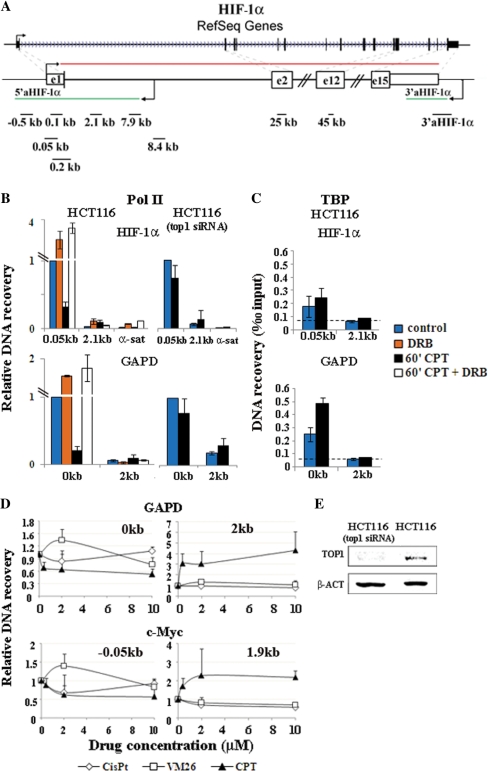
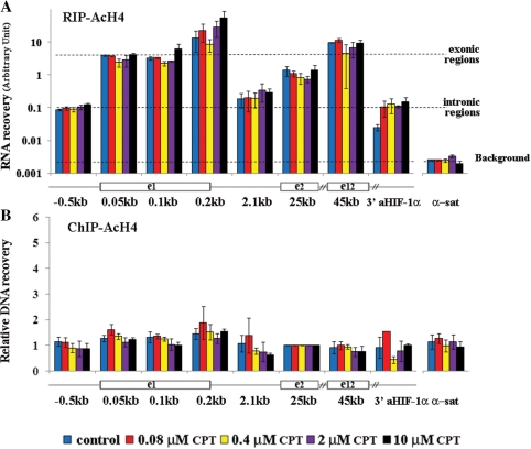
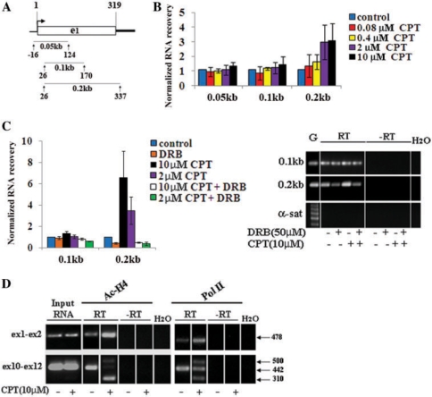
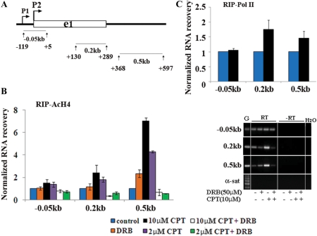
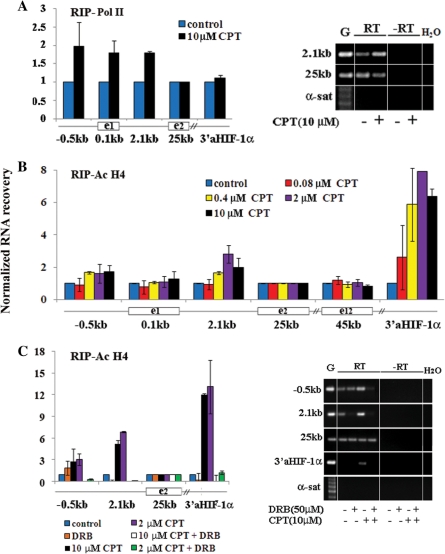
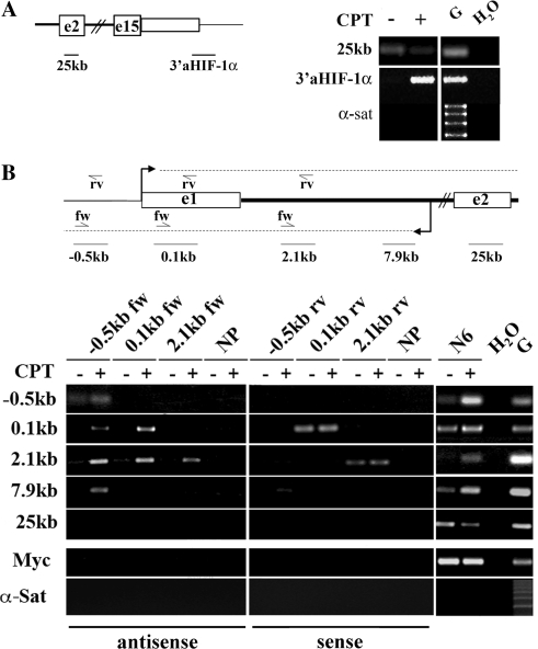
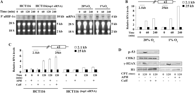
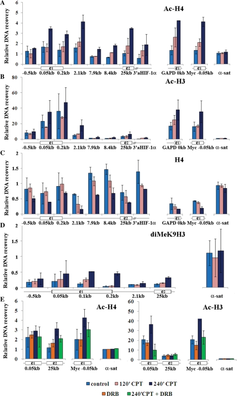
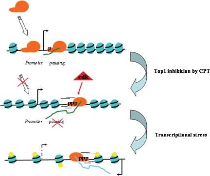
Top1 inhibition by camptothecin (CPT) perturbs RNA polymerase II (Pol II) density at promoters and along transcribed genes suggesting an involvement of Top1 in Pol II pausing. Here, we demonstrate that Top1 inhibition favors Pol II escape from a promoter-proximal pausing site of the human HIF-1alpha gene in living cells. Interestingly, alternative splicing at exon 11 was markedly altered in nascent HIF-1alpha mRNAs, and chromatin structure was also affected with enhanced histone acetylation and reduced nucleosome density in a manner dependent on cdk activity. Moreover, CPT increases transcription of a novel long RNA (5'aHIF1alpha), antisense to human HIF-1alpha mRNA, and a known antisense RNA at the 3'-end of the gene, while decreasing mRNA levels under normoxic and hypoxic conditions. The effects require Top1, but are independent from Top1-induced replicative DNA damage. Chromatin RNA immunoprecipitation results showed that CPT can activate antisense transcription mediated by cyclin-dependent kinase (cdk) activity. Thus, Top1 inhibition can trigger a transcriptional stress, involving antisense transcription and increased chromatin accessibility, which is dependent on cdk activity and deregulated Pol II pausing. A changed balance of antisense transcripts and mRNAs may then lead to altered regulation of HIF-1alpha activity in human cancer cells.


Figures

Figure 1.
Figure 1.
CPT-induced reduction of Pol II density at the HIF-1α gene promoter is dependent on Top1 and Cdk activity. (A) The human HIF-1α gene is indicated with exons (boxes) and introns (lines). Red and green lines show sense and antisense transcripts. Black lines and numbers (distance from mRNA start) indicate the amplicons used in this work. The lower map is not to scale. (B) HCT116 and HCT116(Top1-siRNA) cells were treated for 1 h with CPT (10 µM) and/or DRB (50 µM). DNA recovery was determined at promoter-proximal (0.05 or 0 kb) and -distal regions (2.1 and 2 kb) of HIF-1α and GAPD genes. α-sat, control α-satellite DNA. Values are normalized to promoter-proximal regions in untreated cells, and are means ± SD of at least four determinations from two independent experiments. Mean recovery with non-immune IgG was 0.08. (C) TBP levels at promoters. Values are means ± SD of four determinations from two independent experiments. The dashed line shows recovery levels with non-immune IgG. (D) Effects of CPT, teniposide (VM26) and cis-platin (Cis-Pt) on Pol II density in human MRC5 cells at gene promoters (0 kb), and internal gene regions (2 and 1.9 kb). Values are normalized to untreated cells, and are means ± SD of at least four determinations from two independent experiments. (E) Top1 content of HCT116(Top-siRNA) cells was about 20% of that of HCT116 cells by western blotting. β-Actin (β-ACT) is a loading control.
Figure 2.
Figure 2.
Levels of chromatin-associated RNAs at the HIF-1α gene locus. (A) HCT116 cells were treated for 1 h with the indicated CPT concentrations. RIP was performed with Abs against anti acetylated histone H4 (Ac-H4). Pelleted RNA was retrotranscribed with random primers, and RNA recovery was determined with qRT-PCR using specific primers corresponding to the indicated regions along the HIF-1α gene, a diagram of which (not in scale) is shown under the graph. The broken lines indicate average recovery of exonic, intronic and centromeric α-satellite (α-sat) DNA regions. The background level was determined with cDNAs retrotranscribed without primers and was similar to α-sat recovery levels. A representative experiment is shown: values are means ± SD of three determinations. (B) Levels of Ac-H4 along the HIF-1α gene after 1-h CPT treatments. ChIP was performed with either anti Ac-H4 or non-immune Abs. Values are normalized to exon 2, and are means ± SD of four to six determinations from two independent experiments.
Figure 3.
Figure 3.
CPT-induced Top1ccs promote Pol II escape from the HIF-1α promoter pause site. (A) Diagram of the HIF-1α exon 1 region, with lines indicating the studied amplicons. Numbers indicate positions from mRNA transcription start. (B) HCT 116 cells were treated for 1 h with CPT, and RIP was performed with anti-acetylated histone H4 Abs (Ac-H4). (C) CPT-induced transcription downstream to the pause site is dependent on Cdk activity. On the right: PCR products of RIP samples. Controls are: genomic DNA (G), no reverse transcription (–RT) and water. α-satellite DNA (α-sat) reactions show no genomic DNA contamination. A representative experiment is reported. (D) Top1cc effects on transcription elongation and alternative splicing of HIF-1α mRNA. Transcription levels at two downstream regions from the promoter pausing site spanning exons 1 and 2, and exons 10 to 12. In the latter case, the observed extra band of 310 bases may correspond to a transcript in which exon 11 has been spliced out. –RT indicates no retrotranscription. In all panels, RNA recovery values, normalized to exon 2 and control samples, are means ± SD of at least four determinations from two independent experiments.
Figure 4.
Figure 4.
CPT-induced Top1ccs promote Pol II escape from the c-Myc P2 promoter. (A) Diagram of the studied c-Myc exon 1 region, with lines indicating the studied amplicons. Numbers indicate positions from P2 transcription start. (B) CPT-induced transcription downstream to the P2 pause site is dependent on Cdk activity. HCT116 cells were treated for 1 h with CPT and/or DRB. RIP was performed with Ab against acH4 histone (Ac-H4) or Pol II (Pol II). The gel shows PCR products of RIP samples. Controls are: genomic DNA (G), no reverse transcription (–RT) and water. α-satellite DNA (α-sat) reactions show no genomic DNA contamination. A representative experiment is reported. (C) RIP was performed with Ab against Pol II. All values are means ± S.D. of four determinations from two independent experiments.
Figure 5.
Figure 5.
Levels of specific nascent RNAs are increased by CPT-induced Top1ccs. HCT116 cells were treated for 1 h with CPT and/or DRB as indicated. Immunoprecipitated RNA was retrotranscribed with random primers, and measured with qRT-PCR. Levels of nascent RNAs at HIF-1α gene locus by using Abs anti Pol II (Pol II) are shown in (A) or anti-acetylated histone H4 (Ac-H4) in (B). (C) Activation of nascent RNAs is dependent on cdk activity as shown by RIP experiments performed with anti Ac-H4 Abs. On the right of (A) and (C), a representative gel of PCR products. Samples are: genomic DNA (G), RIP samples (RT). –RT indicates no reverse transcription. α-sat amplicon shows no genomic DNA contamination. In all panels, values are normalized to exon 2 (25 kb) and control samples, and are means ± SD of four to six determinations from two independent experiments.
Figure 6.
Figure 6.
Antisense transcription at the HIF-1α gene locus is increased by Top1cc. Total RNA was extracted from cells untreated (−) or treated with 10 µM CPT for 4 h (+). (A) PCR analysis was performed with primers as indicated in the map. (B) Upper panel: map of the HIF-1α gene region analyzed (not to scale). Dashed lines show the antisense transcript detected at the 5′-end and the mRNA of the HIF-1α gene. Arrows indicate specific primers used for retrotranscription: rv primers target the mRNA, and fw primers target antisense transcripts. Black lines and numbers indicate regions analyzed by PCR. Lower panel: PCR analyses on primer-specific cDNAs from HCT116 cells. Primers used for retrotranscription are indicated on top of the gel, and amplicons used for PCR analyses are indicated on the left of the gel. Negative and positive controls of retrotranscription were no primers (NP) and random primers (N6), respectively. Negative and positive controls of PCR were water (H2O) and genomic DNA (G), respectively. α-satellite DNA (α-sat) shows no genomic DNA contamination.
Figure 7.
Figure 7.
CPT activation of antisense transcription requires Top1 and is independent from replicative DNA damage. (A) Left panel: levels of 5′aHIF1α antisense RNA in cells treated with 10 μM CPT for the indicated time by northern blots. The probe was a 40-nt oligomer corresponding to the 2 kb amplicon and complementary to antisense RNA. Right panel: levels of HIF-1α mRNA in Jurkat cells treated with 10 μM CPT by northern blots of total RNAs with an exon 2 probe. Agarose staining was to check for equal RNA loading and integrity. (B) Increase of the 5′aHIF-1α transcript (2.1 kb amplicon) in Jurkat cells treated with 10 µM CPT in normoxia (20%) or hypoxia (1%). No DNA contamination was detected as established with α-sat. A representative experiment is shown. (C) Total RNA was extracted from untreated cells or cells treated with 10 µM CPT for 120 min with or without aphidicolin (APH) or caffeine (Caff). Left and right panels correspond to HCT116 and HCT116(top1-siRNA) cells, respectively. In all graphs, RNA levels were normalized to mRNA exon 2 (25 kb) and untreated samples, and the values are means ± SD of four determinations from two independent experiments. (D) Western blots of p53 and γ-H2AX after CPT treatments of HCT116 cells with or without Caff or APH. Total cellular proteins (upper two panels) or histones (lower panels) were analyzed by western blots. CHK2 and H1 histone were used as loading controls.
Figure 8.
Figure 8.
Late effects of CPT on chromatin structure. (A) and (B) HCT116 cells were treated with 10 µM CPT for 2 and 4 h. ChIP was with Abs against acetyl -K5,-K8,-K12 and K16 of histone H4 or acetyl –K9 and –K14 of histone H3. Histone acetylation levels were determined along the HIF-1α gene, GAPD promoter (GAPD 0 kb) and c-Myc P2 promoter (Myc −0.05 kb). Mean DNA recovery with non-immune IgG was 0.41. Values are means ± SD of four to eight determinations from two to four independent experiments. (C) Levels of total histone H4 bound to chromatin. (D) Levels of dimetil -K9 of histone H3 after CPT treatments. A representative ChIP experiment is shown, and values are means ± SD of two to three determinations. Mean DNA recovery with non-immune IgG is 0.59 (E) HCT116 cells were treated with CPT and/or DRB for 4 h. Values are means ± SD of four to six determinations of two independent experiments. In all panels, the values are normalized to the recoveries of α-sat DNA, as the internal standard, and to that of total histone H4 (shown in C).
Figure 9.
Figure 9.
A model of the molecular mechanism triggered by Top1ccs induced by CPT at transcribed genes. Arrows indicate transcription start sites and direction. The blue spheres are nucleosomes and small yellow spheres are acetylation marks. The orange object is Pol II. Green and light blue lines are sense and antisense transcripts, respectively.

References

    1. Champoux JJ. DNA topoisomerases: structure, function, and mechanism. Annu. Rev. Biochem. 2001;70:369–413. - PubMed
    1. Wang JC. Cellular roles of DNA topoisomerases: a molecular perspective. Nat. Rev. Mol. Cell Biol. 2002;3:430–440. - PubMed
    1. Pommier Y. Topoisomerase I inhibitors: camptothecins and beyond. Nat. Rev. Cancer. 2006;6:789–802. - PubMed
    1. Khobta A, Ferri F, Lotito L, Montecucco A, Rossi R, Capranico G. Early effects of topoisomerase I inhibition on RNA polymerase II along transcribed genes in human cells. J. Mol. Biol. 2006;357:127–138. - PubMed
    1. Merino A, Madden KR, Lane WS, Champoux JJ, Reinberg D. DNA topoisomerase I is involved in both repression and activation of transcription. Nature. 1993;365:227–232. - PubMed

Publication types