. 2009 Nov;136(22):3741-5.
doi: 10.1242/dev.037317.

The Id2+ distal tip lung epithelium contains individual multipotent embryonic progenitor cells

Affiliations

The Id2+ distal tip lung epithelium contains individual multipotent embryonic progenitor cells

Emma L Rawlins et al. Development. 2009 Nov.

Abstract

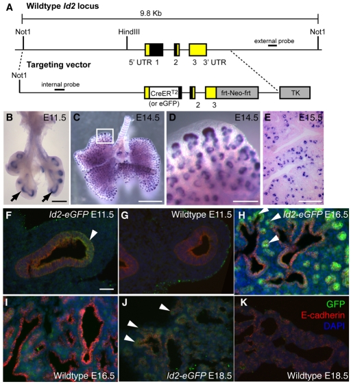
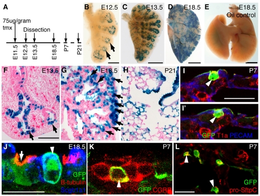
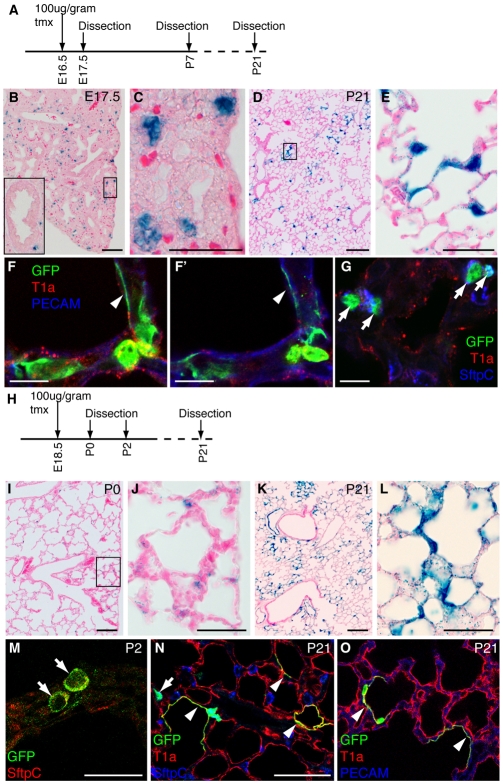
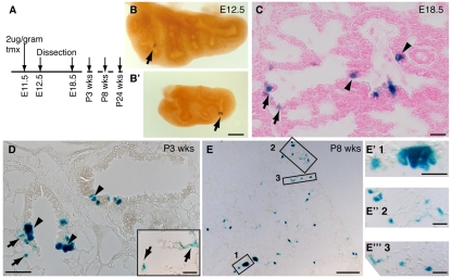
The conducting airways (bronchi and bronchioles) and peripheral gas exchange (alveolar) regions of the mammalian lung are generated by a process of branching morphogenesis. Evidence suggests that during embryonic development, the undifferentiated epithelial progenitors are located at the distal tips of the branching epithelium. To test this hypothesis, we used an Id2-CreER(T2) knock-in mouse strain to lineage trace the distal epithelial tip cells during either the pseudoglandular or canalicular phases of development. During the pseudoglandular stage, the tip cells both self-renew and contribute descendents to all epithelial cell lineages, including neuroendocrine cells. In addition, individual Id2(+) tip cells can self-renew and contribute descendents to both the bronchiolar and alveolar compartments. By contrast, during the later canalicular stage, the distal epithelial tip cells only contribute descendents to the alveoli. Taken together, this evidence supports a model in which the distal tip of the developing lung contains a multipotent epithelial population, the fate of which changes during development.


Figures

Fig. 1.
Fig. 1.
Id2 expression and gene targeting. (A) Targeting vectors. The majority of the mouse Id2 coding region was replaced with a CreERT2 or eGFP cassette. The PGK-Neo cassette (frt-Neo-frt) was removed by FLP-mediated excision after germline transmission (Farley et al., 2000). TK, PGK-thymidine kinase. (B-E) In situ hybridization for Id2 mRNA. (B) E11.5; (C) E14.5; (D) Higher magnification of box in C. (E) E15.5. Id2 is highly expressed in tip epithelium (arrows). (F-K) Id2-eGFP (F,H,J) and wild-type (G,I,K) lungs. (F,G) E11.5; (H,I) E16.5; (J,K) E18.5. Green, anti-GFP; red, anti-E-cadherin (epithelium); blue, DAPI (nuclei). Arrowheads, eGFP+ epithelial tips. Scale bars: 0.25 cm in B; 0.5 cm in C; 200 μm in D,F-K.
Fig. 2.
Fig. 2.
Id2+ tip epithelial cells labeled at the pseudoglandular stage contribute descendents to all lineages. (A) Id2-CreERT2; Rosa26R-lacZ or Rosa26R-eYFP mouse embryos were exposed to tmx at E11.5 and sacrificed at intervals. (B-E) X-Gal stained (blue) lungs. (B) E12.5. Epithelial staining is restricted to tips (arrows). Rare mesenchymal cells are also labeled, consistent with a low level of Id2 expression in this population. (C) E13.5. (D) E18.5. Note the staining throughout the epithelium. (E) E18.5 vehicle-only control showing no tmx-independent recombination. (F-H) X-Gal-stained (blue) lung sections. (F) E13.5. (G) E18.5. Staining is prominent in the tip epithelium (arrows). (H) P21. Staining is seen throughout the bronchioles and alveoli. (I,I′) P7. Two optical sections from a confocal stack. Green, anti-GFP, lineage label; red, anti-T1α, type 1 and 2 cells at P7 (see Fig. S2 in the supplementary material); blue, anti-Pecam, endothelium. Arrowhead, labeled type 1 cell, based on morphology and position adjacent to endothelial cell. (J) E18.5 confocal maximum projection. Green, anti-GFP, lineage label; red, anti-β-tubulin, ciliated cells; blue, anti-Scgb1a1, Clara cells. Arrowhead, lineage-labeled Clara cell. Arrow, lineage-labeled ciliated cell. (K) P7 confocal maximum projection. Green, anti-GFP, lineage label; red, anti-CGRP (Calca), neuroendocrine (NE) cells. Arrowhead, lineage-labeled NE cell (n=24 neuroendocrine bodies with one or more GFP+ cells, five lungs scored). (L) P7 confocal maximum projection. Green, anti-GFP, lineage label; red, anti-pro-SftpC, type 2 cells. Arrowheads, lineage-labeled type 2 cells. Scale bars: 1.5 mm in B; 3 mm in C; 6 mm in D; 7.5 mm in E; 500 μm in F-H; 20 μm in I-L.
Fig. 3.
Fig. 3.
Id2+ epithelial tip cells labeled at later stages only contribute descendents to the alveoli. (A) Time course of experiment shown in B-G. Id2-CreERT2; Rosa26R-lacZ or Rosa26R-eYFP mouse embryos were exposed to tmx at E16.5 and sacrificed at intervals. (B-E) X-Gal-stained (blue) lung sections. (B) E17.5. Note staining in epithelial tips and not in the more proximal airways (inset). (C) Higher magnification of box in B showing labeled tip cells. (D) P21. Note labeling throughout the alveoli. (E) Higher magnification of box in D showing lineage-labeled type 1 and 2 cells. (F-G). P7 sections. (F,F′) Two confocal sections from same z-stack showing lineage-labeled type 1 cell (arrowhead) tightly apposed to, but separate from, the endothelium. Green, anti-GFP, lineage label; red, anti-T1α, type 1 and 2 cells (see Fig. S2 in the supplementary material); blue, anti-Pecam, endothelium. (G) Lineage-labeled type 2 cells (arrows). Green, anti-GFP, lineage label; red, anti-T1α, type 1 and 2 cells; blue, anti-pro-SftpC, type 2 cells. (H) Time course of experiment in I-O. Id2-CreERT2; Rosa26R-lacZ or Rosa26R-eYFP embryos were exposed to tmx at E18.5 and sacrificed at intervals. (I-L) X-Gal-stained (blue) lung sections. (I) P0. Labeling in alveoli. (J) Higher magnification of box in I showing labeling restricted to type 2 cells and a small number of endothelial cells. (K) P21. Labeling throughout the alveoli. (L) Higher magnification of K, lineage-labeled type 1 and 2 cells. (M) P2. Lineage-labeled type 2 cells (arrows). Green, anti-GFP, lineage label; red, anti-pro-SftpC, type 2 cells. (N) P21. Lineage-labeled type 1 (arrowheads) and type 2 (arrow) cells. Green, anti-GFP, lineage label; red, anti-T1α, type 1 cells; blue, anti-pro-SftpC, type 2 cells. (O) P21. Lineage-labeled type 1 cells (arrowheads). Green, anti-GFP, lineage label; red, anti-T1α, type 1 cells; blue, anti-Pecam, endothelium. Scale bars: 50 μm in B-E,N,O; 10 μm in F-G; 1 mm in I,K; 20 μm in J,L,M.
Fig. 4.
Fig. 4.
Single Id2+ tip epithelial cells can give rise to both bronchiolar and alveolar lineages. (A) Id2-CreERT2; Rosa26R-lacZ mouse embryos were exposed to a low dose of tmx at E11.5 and sacrificed at intervals. (B,B′) E12.5 wholemount lungs showing individual clones (arrows). Pairs of cells are frequently seen, suggesting that lineage-labeled cells divided prior to dissection. (C) E18.5 section. Note lineage-labeled bronchiolar (arrowheads) and alveolar (arrows) cells. (D) Lung section from a 3-week-old mouse. Note the larger groups of lineage-labeled cells. Inset shows labeled alveolar cells in the adjacent section. (E-E‴) Lung section from an 8-week-old mouse. Low-magnification image showing clone size. Box 1 shows a bronchiolar clone at higher magnification. Boxes 2 and 3 are high magnifications of alveolar regions showing labeled type 1 and 2 cells. Scale bars: 1 mm in B,E; 20 μm in C and insets in D,E′-E‴; 40 μm D.

References

    1. Cardoso, W. V. and Lu, J. (2006). Regulation of early lung morphogenesis: questions, facts and controversies. Development 133, 1611-1624. - PubMed
    1. Eblaghie, M. C., Reedy, M., Oliver, T., Mishina, Y. and Hogan, B. L. (2006). Evidence that autocrine signaling through Bmpr1a regulates the proliferation, survival and morphogenetic behavior of distal lung epithelial cells. Dev. Biol. 291, 67-82. - PubMed
    1. Farley, F. W., Soriano, P., Steffen, L. S. and Dymecki, S. M. (2000). Widespread recombinase expression using FLPeR (flipper) mice. Genesis 28, 106-110. - PubMed
    1. Hayashi, S. and McMahon, A. P. (2002). Efficient recombination in diverse tissues by a tamoxifen-inducible form of Cre: a tool for temporally regulated gene activation/inactivation in the mouse. Dev. Biol. 244, 305-318. - PubMed
    1. Hong, K. U., Reynolds, S. D., Giangreco, A., Hurley, C. M. and Stripp, B. R. (2001). Clara cell secretory protein-expressing cells of the airway neuroepithelial body microenvironment include a label-retaining subset and are critical for epithelial renewal after progenitor cell depletion. Am. J. Respir. Cell Mol. Biol. 24, 671-681. - PubMed

Publication types

MeSH terms

Substances