

Fas ligand expression on T cells is sufficient to prevent prolonged airway inflammation in a murine model of asthma
- PMID: 19855087
- PMCID: PMC2933550
- DOI: 10.1165/rcmb.2008-0454OC
Fas ligand expression on T cells is sufficient to prevent prolonged airway inflammation in a murine model of asthma
Abstract
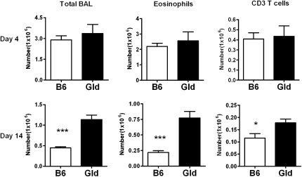
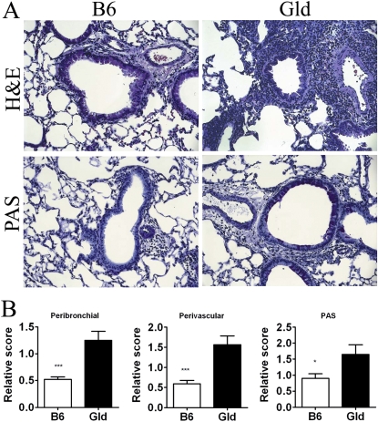
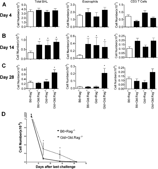
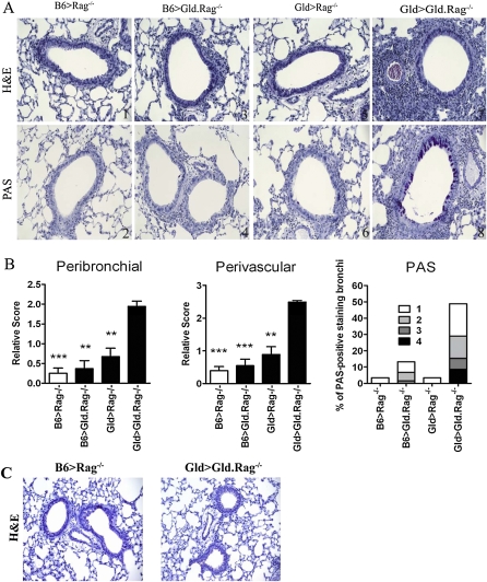
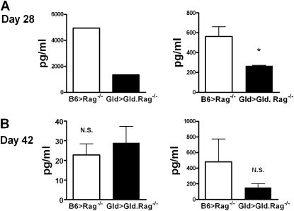
Our previous studies revealed that, in a murine model of asthma, mice that received Fas-deficient T cells developed a prolonged phase of airway inflammation, mucus production, and airway hyperreactivity that failed to resolve even 6 weeks after the last challenge. To investigate how Fas-Fas ligand (FasL) interaction occurs between T cells and other cells in vivo, Gld mice with abnormalities of the FasL signaling pathway were used. The reconstituted mice were made by transferring T cells from B6 or Gld mice to Rag(-/-) or FasL-deficient Rag(-/-) mice. We found that Rag(-/-) mice that received B6 T cells resolved the airway inflammation, whereas FasL-deficient Rag(-/-) mice that received Gld T cells developed a prolonged airway inflammation at Day 28, with decreased IFN-gamma production. Both FasL-deficient Rag(-/-) mice that received B6 T cells and Rag(-/-) mice that received Gld T cells also had completely resolved their airway inflammation by Day 28 after challenge. Interestingly, FasL-deficient Rag(-/-) mice that received Gld T cells eventually resolved airway inflammation at Day 42, with a similar level of IFN-gamma production to that of control group. These results demonstrate that FasL expression on either T cells only or non-T cells only was sufficient for the eventual resolution of airway inflammation, and the prolonged airway inflammation in FasL-deficient Rag(-/-) mice that received Gld T cells was correlated with decreased IFN-gamma production by Gld T cells.
Figures
Similar articles
-
Fas-positive T cells regulate the resolution of airway inflammation in a murine model of asthma.J Exp Med. 2006 May 15;203(5):1173-84. doi: 10.1084/jem.20051680. Epub 2006 Apr 17. J Exp Med. 2006. PMID: 16618792 Free PMC article.
-
Low doses of exogenous interferon-γ attenuated airway inflammation through enhancing Fas/FasL-induced CD4+ T cell apoptosis in a mouse asthma model.J Interferon Cytokine Res. 2012 Nov;32(11):534-41. doi: 10.1089/jir.2012.0016. Epub 2012 Sep 20. J Interferon Cytokine Res. 2012. PMID: 22994871 Free PMC article.
-
Fas-ligand-expressing adenovirus-transfected dendritic cells decrease allergen-specific T cells and airway inflammation in a murine model of asthma.J Mol Med (Berl). 2006 Jul;84(7):595-603. doi: 10.1007/s00109-006-0047-3. Epub 2006 Mar 25. J Mol Med (Berl). 2006. PMID: 16565865
-
Fas ligand (gld)- and Fas (lpr)-deficient mice do not show alterations in the extravasation or apoptosis of inflammatory neutrophils.J Leukoc Biol. 1998 Sep;64(3):373-83. doi: 10.1002/jlb.64.3.373. J Leukoc Biol. 1998. PMID: 9738665
-
Treatment of chronic sialadenitis in a murine model of Sjögren's syndrome by local fasL gene transfer.Arthritis Rheum. 2001 Apr;44(4):964-73. doi: 10.1002/1529-0131(200104)44:4<964::AID-ANR154>3.0.CO;2-5. Arthritis Rheum. 2001. PMID: 11315936
Cited by
-
Obesity Enhances Non-Th2 Airway Inflammation in a Murine Model of Allergic Asthma.Int J Mol Sci. 2024 Jun 4;25(11):6170. doi: 10.3390/ijms25116170. Int J Mol Sci. 2024. PMID: 38892358 Free PMC article.
-
Treatment of allergic asthma: modulation of Th2 cells and their responses.Respir Res. 2011 Aug 25;12(1):114. doi: 10.1186/1465-9921-12-114. Respir Res. 2011. PMID: 21867534 Free PMC article. Review.
-
Non-apoptotic Fas (CD95) Signaling on T Cells Regulates the Resolution of Th2-Mediated Inflammation.Front Immunol. 2018 Nov 1;9:2521. doi: 10.3389/fimmu.2018.02521. eCollection 2018. Front Immunol. 2018. PMID: 30443253 Free PMC article.
-
Systemic FasL neutralization increases eosinophilic inflammation in a mouse model of asthma.Allergy. 2012 Mar;67(3):328-35. doi: 10.1111/j.1398-9995.2011.02763.x. Epub 2011 Dec 17. Allergy. 2012. PMID: 22175699 Free PMC article.
-
Effect of reactive oxygen species generation in rabbit corneal epithelial cells on inflammatory and apoptotic signaling pathways in the presence of high osmotic pressure.PLoS One. 2013 Aug 15;8(8):e72900. doi: 10.1371/journal.pone.0072900. eCollection 2013. PLoS One. 2013. PMID: 23977369 Free PMC article.
References
-
- Cohn L, Elias JA, Chupp GL. Asthma: mechanisms of disease persistence and progression. Annu Rev Immunol 2004;22:789–815. - PubMed
-
- Vignola AM, Chanez P, Campbell AM, Souques F, Lebel B, Enander I, Bousquet J. Airway inflammation in mild intermittent and in persistent asthma. Am J Respir Crit Care Med 1998;157:403–409. - PubMed
-
- Vignola AM, Chanez P, Chiappara G, Siena L, Merendino A, Reina C, Gagliardo R, Profita M, Bousquet J, Bonsignore G. Evaluation of apoptosis of eosinophils, macrophages, and T lymphocytes in mucosal biopsy specimens of patients with asthma and chronic bronchitis. J Allergy Clin Immunol 1999;103:563–573. - PubMed
-
- Jayaraman S, Castro M, O'Sullivan M, Bragdon MJ, Holtzman MJ. Resistance to Fas-mediated T cell apoptosis in asthma. J Immunol 1999;162:1717–1722. - PubMed
Publication types
MeSH terms
Substances
Grants and funding
LinkOut - more resources
Full Text Sources
Medical
Molecular Biology Databases
Research Materials
Miscellaneous
