Review
. 2010 Mar;24(3):475-84.
doi: 10.1210/me.2009-0283. Epub 2009 Oct 23.

Minireview: the melanocortin 2 receptor accessory proteins

Affiliations
Review

Minireview: the melanocortin 2 receptor accessory proteins

Tom R Webb et al. Mol Endocrinol. 2010 Mar.

Abstract

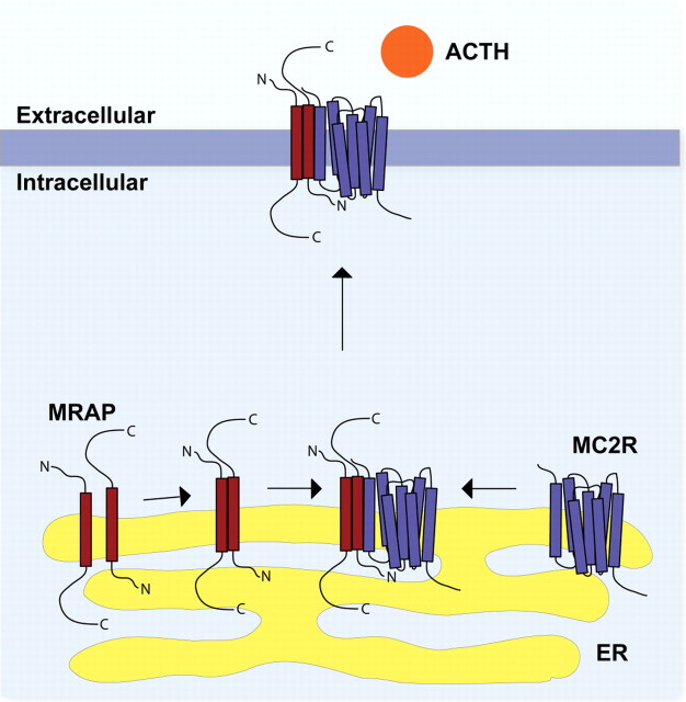
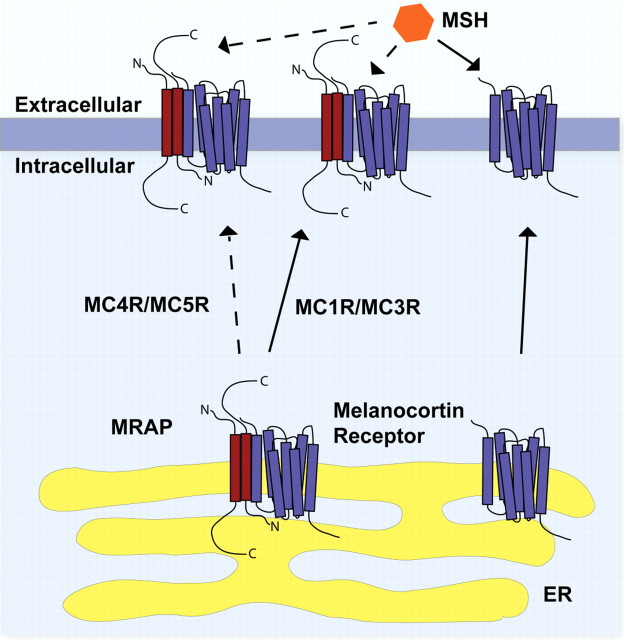
The melanocortin 2 receptor (MC2R) accessory protein, MRAP, is one of a growing number of G protein-coupled receptor accessory proteins that have been identified in recent years that add control and complexity to G protein-coupled receptor functional expression and signal transduction. MRAP interacts directly with MC2R and is essential for its trafficking from the endoplasmic reticulum to the cell surface, where it acts as the receptor for the pituitary hormone ACTH. In addition, MRAP2, a newly described homolog of MRAP, is also able to support the cell surface expression of MC2R. Although it is clear that MRAP is required for MC2R function, the mechanism of MRAP action is only beginning to be understood. Recent work has started to reveal some of these mechanisms and the MRAP domains involved in MC2R functional expression, and new data have shown a potential role for both MRAP and MRAP2 in the regulation of the other melanocortin receptors.


Figures

Fig. 1.
Fig. 1.
Hypothetical model of the interaction of MRAP with the MC2R. MRAP (red) forms an antiparallel homodimer within the ER. This homodimer interacts with the MC2R (purple), possibly assisting its folding into an appropriate conformation. After further posttranslational modification in the ER and Golgi, the heterotrimeric structure then traffics to the cell surface, where it is capable of recognizing and responding to the ACTH peptide.
Fig. 2.
Fig. 2.
Conservation of MRAP and MRAP2. A, MRAP gene structure in different species. The human MRAP gene consists of six exons, with exons 3 to 6 coding for the MRAP peptide. Alternative splicing of exon 4 to 5 or 4 to 6 gives rise to MRAPα and MRAPβ respectively in the human. This gene structure shows surprisingly little conservation between species. Lower mammals (represented by mouse) do not have the noncoding exons 1 and 2, whereas the presence of exon 6 is unclear at present. In lower vertebrates such as zebrafish, MRAP is encoded by three exons, which are homologous to human exons 3 to 5. However, in fugu homologous sequence can only be identified to human exons 3 and 4. B, Sequence alignments of MRAP. Alignment of human MRAPα with MRAPβ and MRAP from Macacca mulatta (marmoset; old world primate), Callicebus moloch (red-bellied titi; new world primate), Bos taurus (domestic cow), Rattus norvegicus (Norway rat), Mus musculus (mouse), Gallus gallus (chicken), Danio zerio (zebrafish), and Takifugu rubripes (fugu). MRAP shows little conservation across species, with sequence homology restricted to the N-terminus and transmembrane regions. Notable conservation lies in the surface expression domain (yellow), including the short region proposed to be essential for ligand binding by MC2R (green), the antiparallel dimerization domain (blue), and the transmembrane domain (pink). There is very little homology over the C-terminus, which varies greatly in length. C, Alignment of human MRAPα with human MRAP2. MRAPα and MRAP2 show 27% sequence identity, although again, the majority of conservation is across the N-terminus and transmembrane regions. D, Alignment of human MRAP2 with mouse MRAP2, Xenopus tropicalis MRAP2, the two zebrafish MRAP2 genes, and fugu MRAP2. As in panel B, the conserved transmembrane domain is highlighted in pink and the predicted antiparallel homodimerization domain is highlighted in blue. bt, B. taurus; camo, C. moloch; dr, zebrafish; gg, chicken; hs, H. sapiens; mamu, M. mulatta; mm, mouse; rn, R. norvegicus; tr, fugu; xtr, X. tropicalis.
Fig. 2.
Fig. 2.
Conservation of MRAP and MRAP2. A, MRAP gene structure in different species. The human MRAP gene consists of six exons, with exons 3 to 6 coding for the MRAP peptide. Alternative splicing of exon 4 to 5 or 4 to 6 gives rise to MRAPα and MRAPβ respectively in the human. This gene structure shows surprisingly little conservation between species. Lower mammals (represented by mouse) do not have the noncoding exons 1 and 2, whereas the presence of exon 6 is unclear at present. In lower vertebrates such as zebrafish, MRAP is encoded by three exons, which are homologous to human exons 3 to 5. However, in fugu homologous sequence can only be identified to human exons 3 and 4. B, Sequence alignments of MRAP. Alignment of human MRAPα with MRAPβ and MRAP from Macacca mulatta (marmoset; old world primate), Callicebus moloch (red-bellied titi; new world primate), Bos taurus (domestic cow), Rattus norvegicus (Norway rat), Mus musculus (mouse), Gallus gallus (chicken), Danio zerio (zebrafish), and Takifugu rubripes (fugu). MRAP shows little conservation across species, with sequence homology restricted to the N-terminus and transmembrane regions. Notable conservation lies in the surface expression domain (yellow), including the short region proposed to be essential for ligand binding by MC2R (green), the antiparallel dimerization domain (blue), and the transmembrane domain (pink). There is very little homology over the C-terminus, which varies greatly in length. C, Alignment of human MRAPα with human MRAP2. MRAPα and MRAP2 show 27% sequence identity, although again, the majority of conservation is across the N-terminus and transmembrane regions. D, Alignment of human MRAP2 with mouse MRAP2, Xenopus tropicalis MRAP2, the two zebrafish MRAP2 genes, and fugu MRAP2. As in panel B, the conserved transmembrane domain is highlighted in pink and the predicted antiparallel homodimerization domain is highlighted in blue. bt, B. taurus; camo, C. moloch; dr, zebrafish; gg, chicken; hs, H. sapiens; mamu, M. mulatta; mm, mouse; rn, R. norvegicus; tr, fugu; xtr, X. tropicalis.
Fig. 3.
Fig. 3.
Functional domains of MRAP. Three functionally distinct domains of MRAP can be defined. The transmembrane domain (red; residues 36-61) is responsible for membrane anchorage of MRAP and for the physical interaction between MRAP and MC2R and between MRAP and its homodimeric partner. The cell-surface expression domain (yellow; residues 9-24) was shown by Webb et al. (26 ) to be required for effective trafficking of the MC2R to the plasma membrane. Sebag and Hinkle (27 ) reported that the four-amino-acid sequence LDYI between positions 18 and 21 (green) was required for effective MC2R ligand binding and signal transduction. The short sequence between positions 31 and 37 immediately N-terminal to the transmembrane (blue) domain was found to be required for adoption of the antiparallel homodimer structure unique to these proteins (27 ).
Fig. 4.
Fig. 4.
Hypothetical model of the regulation of MCRs by the MRAP proteins. In the absence of MRAP (or MRAP2), the MCRs (purple) traffic to the cell surface from the ER and respond to MSH peptides. In a heterotrimeric complex with MRAP or MRAP2 (red), both MC4R and MC5R show impaired cell surface expression. MRAP or MRAP2 does not change the trafficking of MC1R and MC3R. At the cell surface, these heterotrimeric MRAP-MCR complexes show reduced signaling in response MSH.

References

    1. Metherell LA, Chapple JP, Cooray S, David A, Becker C, Rüschendorf F, Naville D, Begeot M, Khoo B, Nürnberg P, Huebner A, Cheetham ME, Clark AJL2005. Mutations in MRAP, encoding a new interacting partner of the ACTH receptor, cause familial glucocorticoid deficiency type 2. Nat Genet 37:166–170 - PubMed
    1. Cone RD, Mountjoy KG, Robbins LS Nadeau JH, Johnson KR, Roselli-Rehfuss L, Mortrud MT1993. Cloning and functional characterization of a family of receptors for the melanotropic peptides. Ann NY Acad Sci 680:342–363 - PubMed
    1. Mountjoy KG, Robbins LS, Mortrud MT, Cone RD1992. The cloning of a family of genes that encode the melanocortin receptors. Science 257:1248–1251 - PubMed
    1. Cone RD2006. Studies on the physiological functions of the melanocortin system. Endocr Rev 27:736–749 - PubMed
    1. Noon LA, Franklin JM, King PJ, Goulding NJ, Hunyady L, Clark AJ2002. Failed export of the adrenocorticotrophin receptor from the endoplasmic reticulum in non-adrenal cells: evidence in support of a requirement for a specific adrenal accessory factor. J Endocrinol 174:17–25 - PubMed

MeSH terms