

Optic nerve head (ONH) topographic analysis by stratus OCT in normal subjects: correlation to disc size, age, and ethnicity
- PMID: 19855299
- PMCID: PMC3417149
- DOI: 10.1097/IJG.0b013e3181b6e5cd
Optic nerve head (ONH) topographic analysis by stratus OCT in normal subjects: correlation to disc size, age, and ethnicity
Abstract
Purpose: To study optic nerve head (ONH) topography parameters measured by Stratus optical coherence tomography (OCT) in normal subjects and to analyze ONH data for differences in relation to disc size, ethnicity, and age.
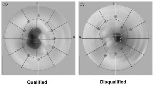
Methods: Three hundred sixty-seven normal subjects underwent Stratus optical coherence tomography ONH measurement using the fast optic disc scan protocol software package 3.0. Only ONH scans meeting specific qualification criteria were included for data analysis ensuring appropriate scan quality and reliability. ONH topographic parameters of qualified scans were analyzed for differences in regards to optic disc size, age, and ethnicity.
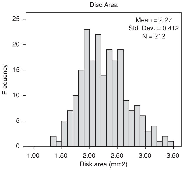
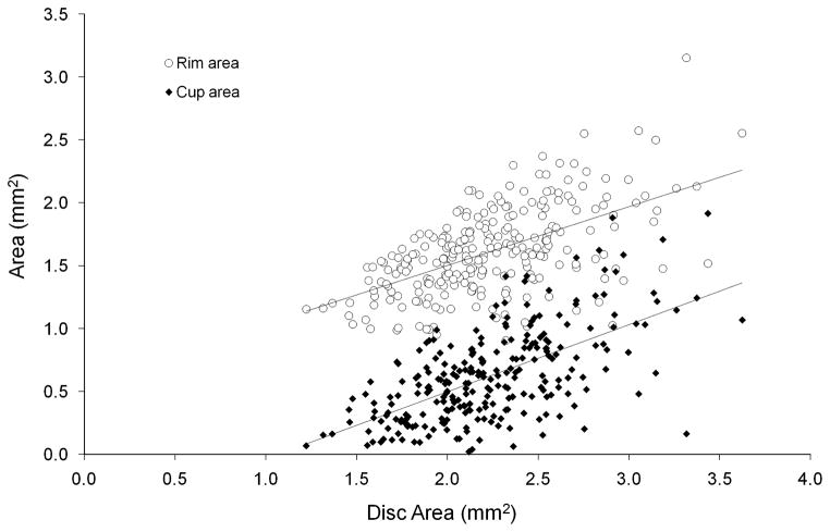
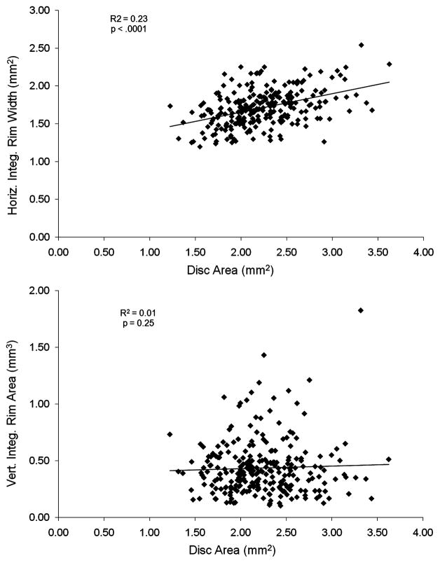
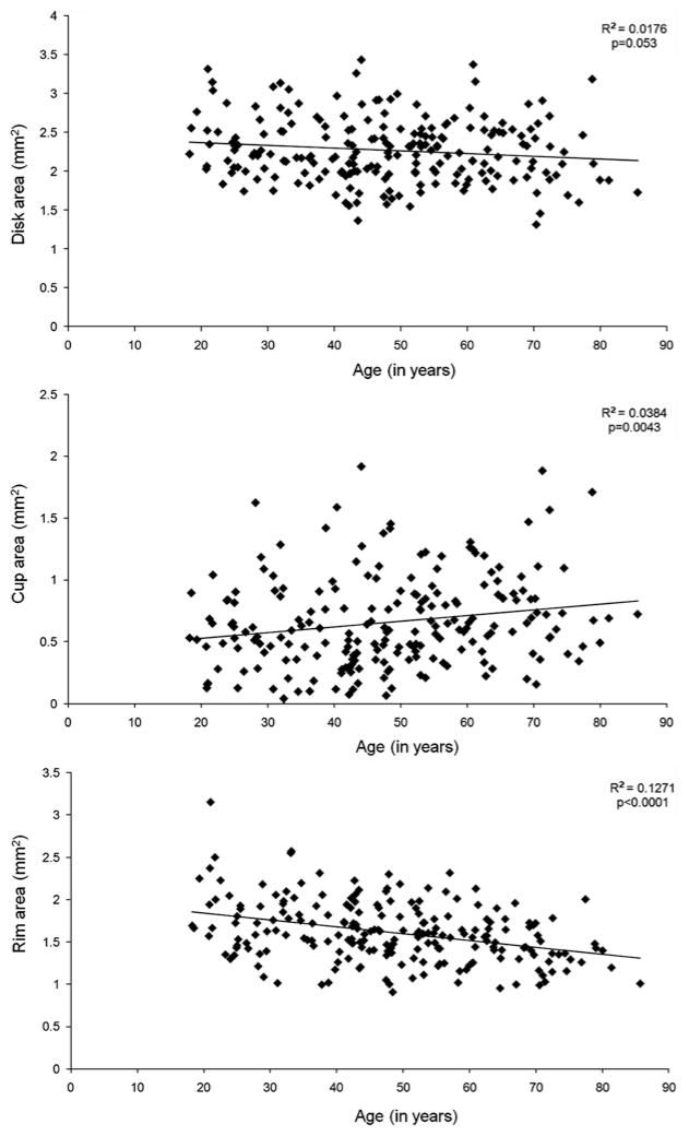
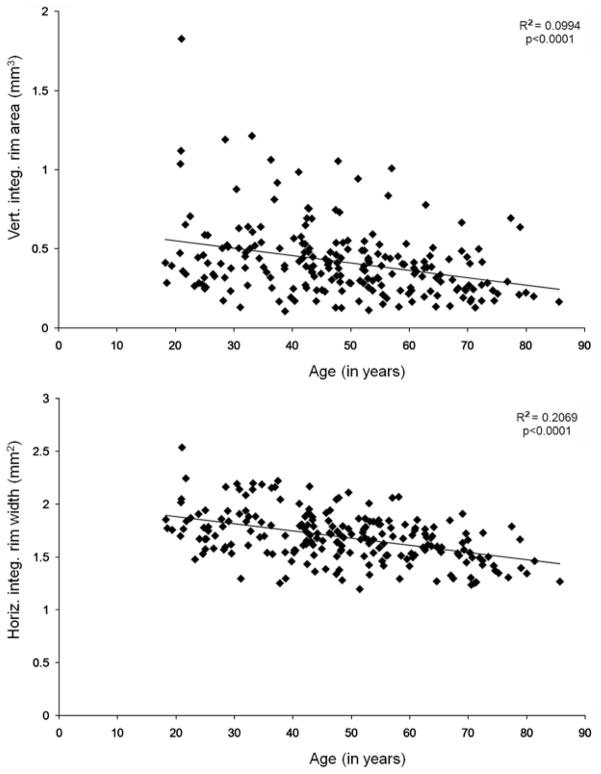
Results: Two hundred and twelve qualified ONH scans were included for data analysis. Mean disc area was 2.27+/-0.41 mm and optic cup area, rim area, and horizontal integrated rim width increased with disc size, whereas vertical integrated rim area did not. Vertical integrated rim area, horizontal integrated rim width, and rim area decreased and cup area increased with age. Mean optic disc area was larger in African-Americans as compared with Hispanics or Whites and this difference was statistically significant.
Conclusions: Optic cup area, rim area, and horizontal integrated rim width correlated to disc size. Vertical integrated rim area, horizontal integrated rim width, rim area, and cup area, changed with age. African-American optic discs had larger disc area measurements as compared with Whites optic discs and this difference was statistically significant.
Figures
References
-
- Leung CK, Cheng AC, Chong KK, et al. Optic disc measurements in myopia with optical coherence tomography and confocal scanning laser ophthalmoscopy. Invest Ophthalmol Vis Sci. 2007;48:3178–1383. - PubMed
-
- Hoffman EM, Bowd C, Medeiros FA, et al. Agreement among 3 optical imaging methods for the assessment of optic disc topography. Ophthalmology. 2005;112:2149–2156. - PubMed
-
- Schuman JS, Wollstein G, Farra T, et al. Comparison of optic nerve head measurements obtained by optical coherence tomography and confocal scanning laser ophthalmoscopy. Am J Ophthalmol. 2003;135:504–512. - PubMed
-
- Olmedo M, Cadarso-Suarez C, Gomez-Ulla F, et al. Reproducibility of optic nerve head measurements obtained by optical coherence tomography. Eur J Ophthalmol. 2005;15:486–492. - PubMed
Publication types
MeSH terms
Grants and funding
LinkOut - more resources
Full Text Sources
Medical
