. 2009 Nov;11(11):1332-9.
doi: 10.1038/ncb1977. Epub 2009 Oct 25.

Intraflagellar transport is required for polarized recycling of the TCR/CD3 complex to the immune synapse

Affiliations

Intraflagellar transport is required for polarized recycling of the TCR/CD3 complex to the immune synapse

Francesca Finetti et al. Nat Cell Biol. 2009 Nov.

Abstract

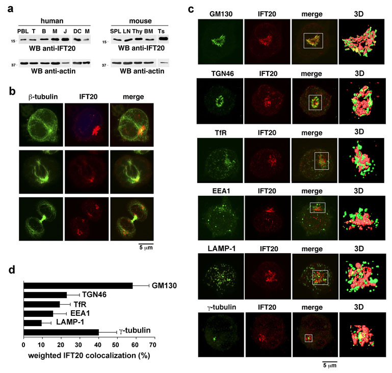
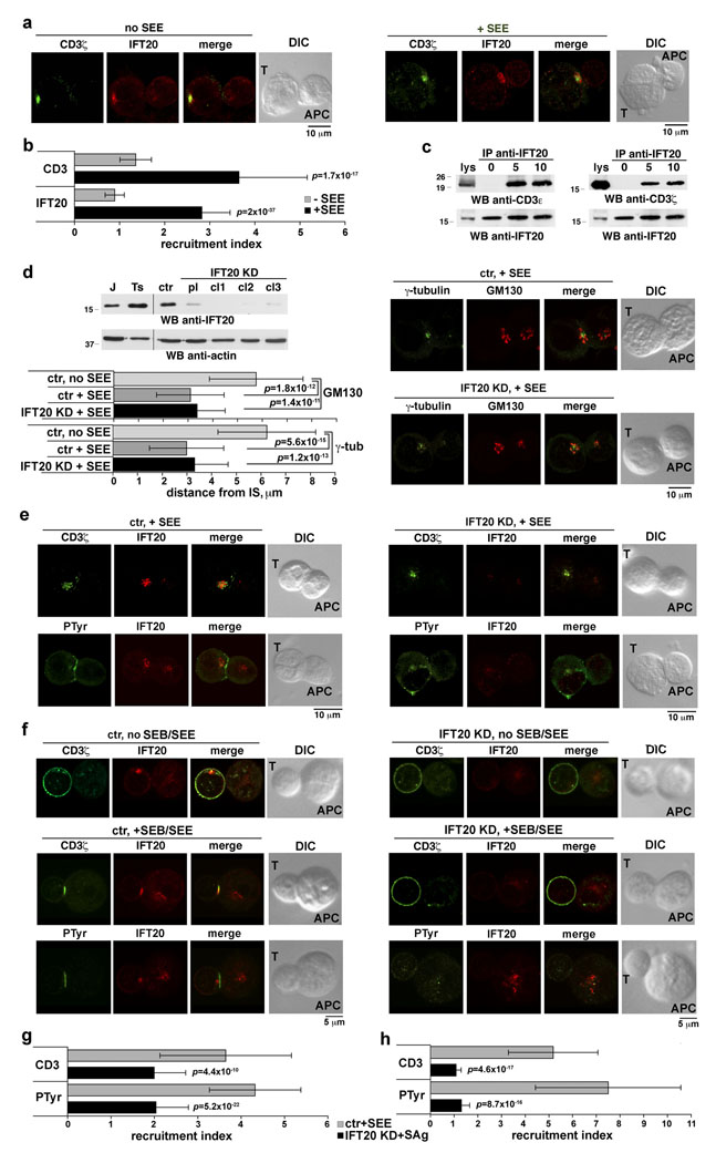
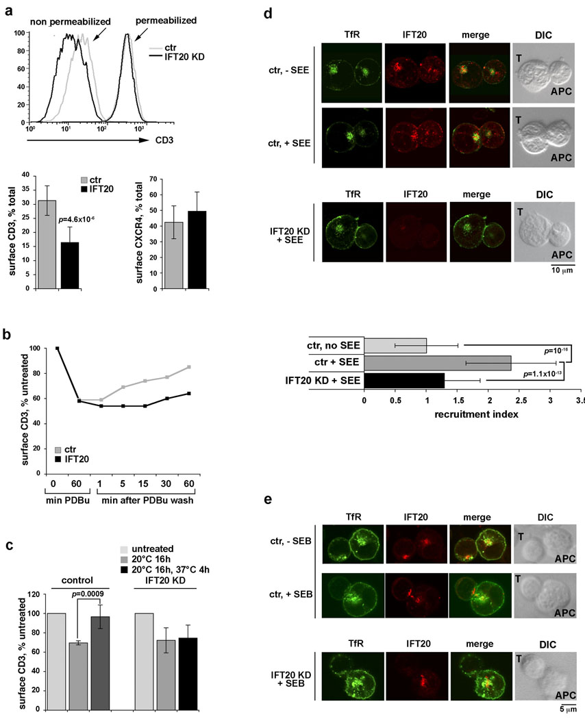
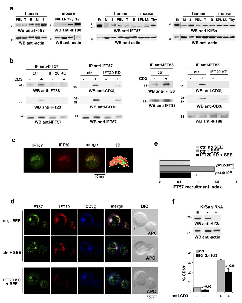
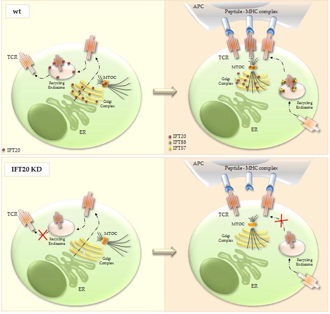
Most eukaryotic cells have a primary cilium which functions as a sensory organelle. Cilia are assembled by intraflagellar transport (IFT), a process mediated by multimeric IFT particles and molecular motors. Here we show that lymphoid and myeloid cells, which lack primary cilia, express IFT proteins. IFT20, an IFT component essential for ciliary assembly, was found to colocalize with both the microtubule organizing centre (MTOC) and Golgi and post-Golgi compartments in T-lymphocytes. In antigen-specific conjugates, IFT20 translocated to the immune synapse. IFT20 knockdown resulted in impaired T-cell receptor/CD3 (TCR/CD3) clustering and signalling at the immune synapse, due to defective polarized recycling. Moreover, IFT20 was required for the inducible assembly of a complex with other IFT components (IFT57 and IFT88) and the TCR. The results identify IFT20 as a new regulator of immune synapse assembly in T cells and provide the first evidence to implicate IFT in membrane trafficking in cells lacking primary cilia, thereby introducing a new perspective on IFT function beyond its role in ciliogenesis.


Figures

Figure 1
Figure 1. IFT20 is expressed in T cells and associates with Golgi apparatus and MTOC
a. Immunoblot analysis of IFT20 in lysates of human and murine lymphoid and myeloid cells. PBL, peripheral blood lymphocytes; T, T-cells; B, B-cells; M, monocytes; J, Jurkat cells; DC, dendritic cells; SPL, spleen; LN, lymph node; Thy, thymus; BM, bone marrow; Ts, testis (positive control). b,c. Immunofluorescence analysis of IFT20 localization in Jurkat cells costained with antibodies against (b) β-tubulin or (c) γ-tubulin or markers of membrane compartments. Panel b shows cells in interphase (top), metaphase (middle) and telophase (bottom). In panel c, GM130, cis-Golgi; TGN46, trans-Golgi network; TfR, recycling endosomes; EEA1, early endosomes; LAMP-1, lysosomes. Z-stack projections are shown. The white box in the merge images represents the field of view for the 3D reconstructions based on sequential 0.5 µm sections shown on the last panel of each row. The corresponding animations are shown in the supplemental online information (Movie 1–Movie 6). d. Quantification of the weighted colocalization (as percentage) of the different subcellular organelles/compartments with the IFT20 staining (mean ±SD). 30 cells were analyzed for each marker. All of the pairwise comparisons of the IFT20 colocalisions with the GM130, TGN46, TfR, EEA1, LAMP-1 and γ-tubulin were statistically significant (p<0.01 at least) with only two exceptions being: TGN46 vs TfR and TfR vs EEA1.
Figure 2
Figure 2. IFT20 is required for TCR/CD3 clustering and signaling at the IS
a. Immunofluorescence analysis of IFT20 and CD3ζ localization in conjugates of Jurkat cells and APC, in the presence or absence of SEE. Median optical sections are shown. b. Relative IFT20 and CD3ζ fluorescence at the T-cell:APC contact site compared to the remaining T-cell membrane (CD3ζ) or T-cell area (IFT20) (expressed as relative recruitment index). Measurements on conjugates from ≥3 independent experiments (n=50). Error bars, SD. c. Immunoblot with anti-CD3ζ and -CD3ε antibodies of IFT20-specific immunoprecipitates from lysates of Jurkat cells either unstimulated or stimulated with anti-CD3 mAb for 5–10 min (whole blot in Fig.S6a). d. Top left, Immunoblot of IFT20 in lysates of Jurkat cells, a Jurkat line transfected with empty vector (ctr), and three Jurkat clones (cl1-3) transfected with constructs encoding IFT20-specific siRNA (KD) (whole blot in Fig.S6b; all samples on the same gel). A pool (pl) of the three IFT20-KD clones (IFT20 reduction in the pool ~75%) was used for subsequent experiments. Right, Immunofluorescence analysis of SEE-specific conjugates of control or IFT20-KD Jurkat cells and APC. Cells were costained with anti-γ-tubulin and -GM130 antibodies. Bottom right, Mean distance±SD of the MTOC and of the central point of the cis-Golgi from the T-cell:APC contact site. Measurements on conjugates from ≥3 independent experiments (n=50). e,f. Immunofluorescence analysis of SEE-specific conjugates of control or IFT20-KD Jurkat cells and APC (e) or of SEB/SEE-specific conjugates using peripheral T-cells transiently transfected with empty vector or the IFT20 RNAi construct (IFT20 reduction ~40%, ~80% and ~70% for donors 1–3, respectively, as assessed by immunoblot) (f). Cells were costained with anti-IFT20 and either anti-CD3ζ or anti- PTyr antibodies. g,h. Relative CD3ζ or PTyr fluorescence at the T-cell:APC contact site compared to the remaining T-cell membrane (CD3ζ) or T-cell area (PTyr) (expressed as relative recruitment index) in Jurkat cells (g) and T-cells from healthy donors (h). Measurements on conjugates from ≥3 independent experiments for Jurkat cells (n=50), or on conjugates from duplicate samples from 3 independent donors for peripheral T-cells (n=30, 10/donor).
Figure 3
Figure 3. IFT20 is required for trafficking of the TCR/CD3 complex in recycling endosomes
a. Representative FACS profiles and relative levels of CD3 in intact (surface CD3) vs permeabilized (total CD3) control/IFT20-KD Jurkat cells (n≥9). Data from similar experiments are shown for CXCR4 (right; n≥5). b. Relative levels of surface CD3 in control/IFT20-KD cells untreated, or treated with PDBu for 1h, or treated with PDBu then washed and incubated at 37°C for the indicated times. A representative experiment is shown (n=4). c. Relative levels of surface CD3 in control/IFT20-KD cells untreated, or incubated at 20°C for 16h, or incubated at 20°C then shifted to 37°C for 4h. Receptor expression was measured by flow cytometry, and calculated on the MFI levels (n=3). Surface CD3 recovery was 88.8% in control vs 8.3% in IFT20-KD (p=0.019). Error bars, SD. d. Immunofluorescence analysis of IFT20 and TfR localization in conjugates of control or IFT20-KD Jurkat cells and APC, either unpulsed or pulsed with SEE. Median optical sections are shown. The graph on the right shows the relative TfR fluorescence at the the T-cell:APC contact site compared to the remaining T-cell area (expressed as relative recruitment index). Error bars, SD. The measurements were taken on ≥50 conjugates from ≥3 independent experiments, respectively. e. Immunofluorescence analysis with anti-TfR and anti-IFT20 antibodies of TfR localization in SEB-specific conjugates using SEB-specific peripheral T-cells from a representative healthy donor transiently transfected with either empty vector or the IFT20 RNAi construct (n=3).
Figure 4
Figure 4. IFT20 inducibly associates with other IFT components in response to TCR triggering
a. Immunoblot analysis of IFT88, IFT57 and Kif3a in human and murine lymphoid and myeloid cell lysates (samples on the same gel). Abbreviations are as in Figure1a. b. Immunoblot analysis with the indicated antibodies of anti-IFT57 (left) or anti-IFT88 (right) specific immunoprecipitates from lysates of Jurkat cells either unstimulated or stimulated with anti-CD3 mAb for 10 min (whole blots in Fig.S6c,d). A mouse testis lysate was included in each gel as control (not shown). c. Immunofluorescence analysis of IFT57 and IFT20 localization in Jurkat cells. A Z-stack projection is shown. The white box in the merge image represents the field of view for the 3D reconstruction based on sequential 0.5 µm sections shown on the last panel. The corresponding animation is shown in the Supplemental Information (Movie 7). Weighted colocalization (as percentage) with the IFT20 staining was 42.0 ±7.4 (n=30). d. Immunofluorescence analysis of IFT57 and IFT20 localization in conjugates of control or IFT20-KD Jurkat cells and APC, in the presence or absence of antigen (SEE). Cells were costained with antibodies against CD3ζ. Median optical sections are shown. e. Relative IFT57 fluorescence at the T-cell:APC contact site compared to the remaining T-cell area (expressed as relative recruitment index). Error bars, SD. Measurements were taken on 30 conjugates for each condition. f. Percentages of CD69+ control or Kif3a-KD Jurkat cells following stimulation by CD3 cross-linking for 8h, as assessed by flow cytometric analysis of surface CD69. The values of duplicate samples from a representative experiment out of 3 are shown. Error bars, SD. An anti-Kif3a immunoblot of lysates of control and Kif3a-KD Jurkat cells is shown (samples on the same gel).
Figure 5
Figure 5. Schematic representation of the localization and function of IFT20 in TCR/CD3 trafficking and IS formation
Top, In resting T cells IFT20, which is associated with the Golgi complex, the microtubule-organizing center (MTOC) and recycling endosomes, regulates constitutive T-cell antigen receptor (TCR) recycling to the cell surface (left). When the TCR is engaged by cognate peptide antigen presented by MHC on the surface of an antigen-presenting cell (APC), IFT20 promotes TCR clustering at the immune synapse through polarized recycling, as a complex with IFT57 and IFT88 (right). Bottom, In the absence of IFT20 both constitutive TCR recycling (left) and polarized TCR recycling to the immune synapse (right) are impaired. Translocation of the Golgi and MTOC to the immune synapse is conversely not affected by the absence of IFT20. ER, endoplasmic reticulum.

References

    1. Scholey JM, Anderson KV. Intraflagellar transport and cilium-based signaling. Cell. 2006;125:439. - PubMed
    1. Rosenbaum JL, Witman GB. Intraflagellar transport. Nat Rev Mol Cell Biol. 2002;3:813. - PubMed
    1. Follit JA, Tuft RA, Fogarty KE, Pazour GJ. The intraflagellar transport protein IFT20 is associated with the Golgi complex and is required for cilia assembly. Mol Biol Cell. 2006;17:3781. - PMC - PubMed
    1. Jonassen JA, San Agustin J, Follit JA, Pazour GJ. Deletion of IFT20 in the mouse kidney causes misorientation of the mitotic spindle and cystic kidney disease. J Cell Biol. 2008;183:377. - PMC - PubMed
    1. Cemerski S, Shaw A. Immune synapses in T-cell activation. Curr Opin Immunol. 2006;18:298. - PubMed

Publication types

MeSH terms