. 2009 Nov;15(11):1318-21.
doi: 10.1038/nm.2053. Epub 2009 Oct 25.

Extracellular histones are major mediators of death in sepsis

Affiliations

Extracellular histones are major mediators of death in sepsis

Jun Xu et al. Nat Med. 2009 Nov.

Abstract

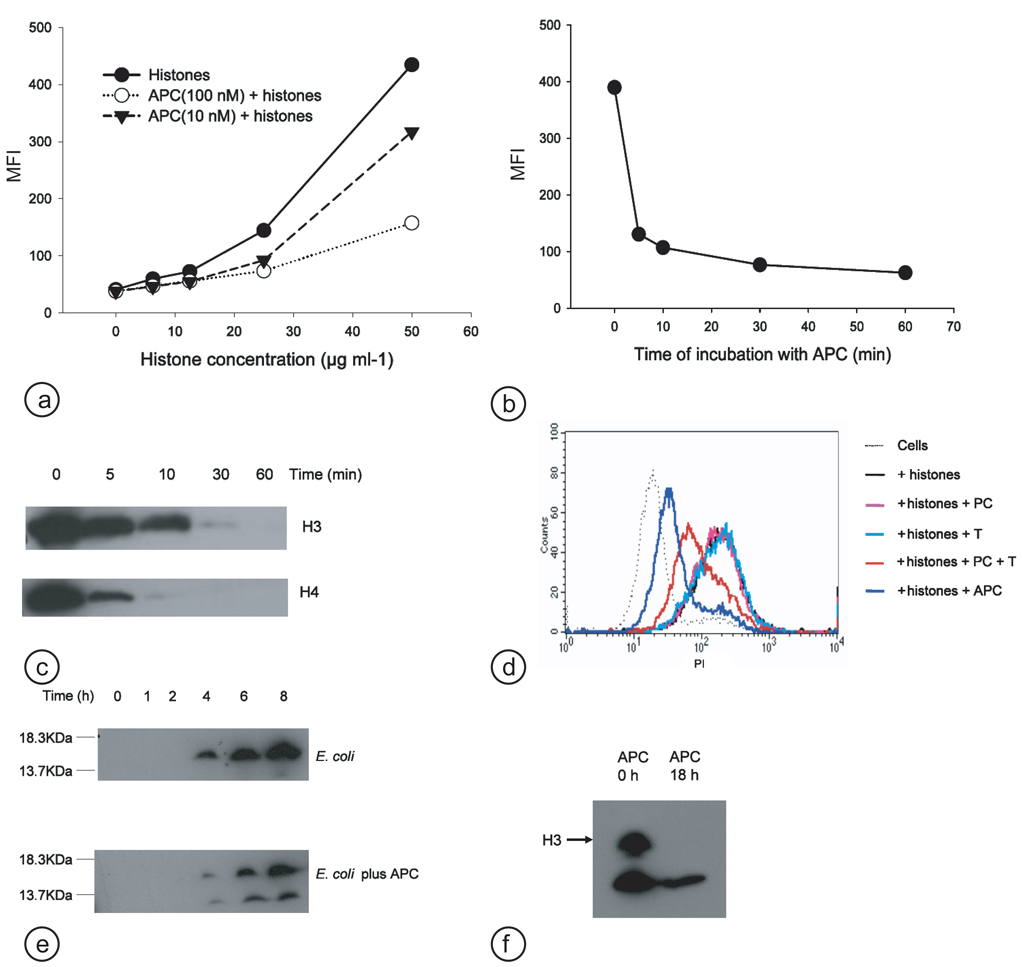
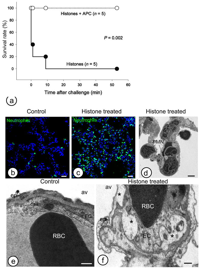
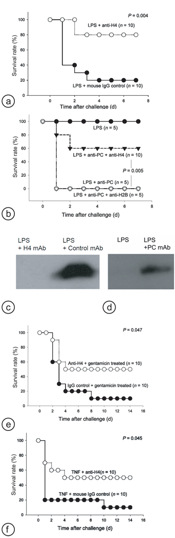
Hyperinflammatory responses can lead to a variety of diseases, including sepsis. We now report that extracellular histones released in response to inflammatory challenge contribute to endothelial dysfunction, organ failure and death during sepsis. They can be targeted pharmacologically by antibody to histone or by activated protein C (APC). Antibody to histone reduced the mortality of mice in lipopolysaccharide (LPS), tumor necrosis factor (TNF) or cecal ligation and puncture models of sepsis. Extracellular histones are cytotoxic toward endothelium in vitro and are lethal in mice. In vivo, histone administration resulted in neutrophil margination, vacuolated endothelium, intra-alveolar hemorrhage and macro- and microvascular thrombosis. We detected histone in the circulation of baboons challenged with Escherichia coli, and the increase in histone levels was accompanied by the onset of renal dysfunction. APC cleaves histones and reduces their cytotoxicity. Co-infusion of APC with E. coli in baboons or histones in mice prevented lethality. Blockade of protein C activation exacerbated sublethal LPS challenge into lethality, which was reversed by treatment with antibody to histone. We conclude that extracellular histones are potential molecular targets for therapeutics for sepsis and other inflammatory diseases.


Figures

Fig.1
Fig.1
Cytotoxicity of extracellular histones toward endothelium and APC cleavage of histones. (a) EA.hy926 cells were cultured with calf thymus histones (50µg ml−1) or calf thymus histone H1, H2A, H2B, H3 or H4 (20µg ml−1) for 1 hr at 37°C. Cell damage was measured by flow cytometry for PI staining. (b) APC (100 nM) was absent or present during the incubations with histones, H3 or H4 in the above assays. (c) SDS–PAGE analysis of purified calf thymus H3 (top panel) or H4 (bottom panel) (100 µg ml−1) incubated with the indicated concentrations of human APC for 1 hr at 37°C. (d) SDS–PAGE analysis of purified calf thymus histone H3 (top panel) or H4 (bottom panel) (100 µg ml−1) incubated with 10 nM human APC in the absence or presence of 0.5 mg ml−1 PS/PC or PE/PS/PC liposomes for 1 hr at 37°C.
Fig.2
Fig.2
APC cleaves histones both in vitro and in vivo. (a) EA.hy926 cells were cultured with calf thymus histones in the indicated concentration in the absence or presence of APC (10 or 100 nM) at 37°C for 1 hr. Cell damage was measured by flow cytometry for PI staining and expressed as mean fluorescence index (MFI). (b) Calf thymus histones (50 µg ml−1) incubated with APC (100 nM) at 37°C for the indicated time and then mixed with PPACK (10 µM) to inactivate APC. The above medium was used to culture EA.hy926 cells for 1 hr for cytotoxicity assay or (c) subjected to SDS–PAGE and Western blotting for H3 or H4 at the times indicated. (d) EA.hy926 cells were cultured with calf thymus histones (50µg ml−1) in the absence or presence of protein C (100 nM), thrombin (T) (10 nM) or APC (100 nM) at 37°C for 30 min. Cell damage was measured by flow cytometry for PI staining. (e) Western blot analysis for H3 of baboon plasma samples at the times indicated after E. coli or E. coli plus APC challenge. (f) Western blot analysis for H3 of plasma samples taken at the times indicated from the start of APC treatment of a human septic patient.
Fig.3
Fig.3
Intravenous injection of histones elicits inflammatory and cell injury responses. (a) Survival rates of mice injected intravenously with calf thymus histones (75 mg per kg) with or without APC (5 mg per kg). (b–d) Pathological changes of mouse lung three hours after intravenous injection of histones (50 mg per kg). Immunofluorescence staining for neutrophil elastase detected massive neutrophil accumulation in the alveolar microvasculature (c: histone treated vs. b: control). Alveolar capillaries are almost fully obstructed by cells, as seen by electron microscopy (d: PMN). (e–f) Histones induce strong alterations of the selective permeability of plasma membranes and subsequent intracellular edema, vacuolization (*) of the intracellular organelles (endoplasmic reticulum, Golgi, and mitochondria), both within endothelial (EC) and type I epithelial cells (ep–I). av, alveolae; cav, caveolae; RBC, red blood cells. Magnification bars: b and c: 50 µm; d, 10 µm; e and f, 500 nm.
Fig.4
Fig.4
Antibody to H4 protects mice from the lethality of LPS, CLP and TNF in vivo. (a) Mice were injected intravenously with a high dose of LPS (10 mg per kg) with antibody to H4 or mouse IgG control antibody (20 mg per kg). Survival rates of each group are indicated. (b) Mice were injected intravenously with a low dose of LPS (1 mg per kg) with or without antibody to protein C (2.5 mg per kg), and with antibody to H4 or H2B (20 mg per kg). Survival rates of each group are indicated. Western blot analysis for H3 of mouse plasma collected (c) 24 h after a high dose of LPS (10 mg per kg) plus antibody to H4 or mouse IgG control antibody (20 mg per kg) or (d) 6 h after a low dose of LPS (1 mg per kg) with or without antibody to protein C (2.5 mg per kg). (e) Survival rates of mice subjected to CLP and then treated with antibody to H4 or mouse IgG control antibody (20 mg per kg) plus gentamicin (5 mg per kg) 6 hr post–CLP. (f) Survival rates of mice injected intravenously with TNF (0.75 mg per kg) plus antibody to H4 or mouse IgG control antibody (5 mg per kg).

Comment in

  • Sepsis: the dark side of histones.
    Chaput C, Zychlinsky A. Chaput C, et al. Nat Med. 2009 Nov;15(11):1245-6. doi: 10.1038/nm1109-1245. Nat Med. 2009. PMID: 19893552 No abstract available.

References

    1. Wang H, et al. HMG–1 as a late mediator of endotoxin lethality in mice. Science. 1999;285:248–251. - PubMed
    1. Bernard GR, et al. Efficacy and safety of recombinant human activated protein C for severe sepsis. N. Engl. J. Med. 2001;344:699–709. - PubMed
    1. Russell JA. Management of sepsis. N. Engl. J. Med. 2006;355:1699–1713. - PubMed
    1. Esmon CT. The protein C pathway. Chest. 2003;124:26S–32S. - PubMed
    1. Kerschen EJ, et al. Endotoxemia and sepsis mortality reduction by non–anticoagulant–activated protein C. J. Exp. Med. 2007;204:2439–2448. - PMC - PubMed

Publication types

MeSH terms