. 2009 Nov;15(11):1259-65.
doi: 10.1038/nm.2036. Epub 2009 Oct 25.

Salmonella disrupts lymph node architecture by TLR4-mediated suppression of homeostatic chemokines

Affiliations

Salmonella disrupts lymph node architecture by TLR4-mediated suppression of homeostatic chemokines

Ashley L St John et al. Nat Med. 2009 Nov.

Abstract

We report that infection of draining lymph nodes (DLNs) by Salmonella typhimurium results in the specific downregulation of the homeostatic chemokines CCL21 and CXCL13, which are essential for normal DLN organization and function. Our data reveal that the mechanism of this suppression is dependent on S. typhimurium LPS (sLPS). The decrease in CCL21 expression involves interaction between sLPS and CCL21-producing cells within DLNs, triggering a distinct Toll-like receptor 4 (TLR4)-mediated host signaling response. In this response, suppressor of cytokine signaling-3 (Socs3) is upregulated, which negatively regulates mothers against decapentaplegic homolog-3 (Smad3)-initiated production of CCL21. Disruption of lymph node architecture and cellular trafficking enhances S. typhimurium virulence and could represent a mechanism of immune suppression used by pathogens that primarily target lymphoid tissue.


Figures

Figure 1
Figure 1
S. typhimurium alters cellular trafficking within DLNs. (a,b) Tissue sections from popliteal DLNs, stained for B cells (B220, green) and T cells (CD3, blue) 24 h after subcutaneous footpad injection of saline or 1 × 10⁵ colony-forming units (CFU) of S. typhimurium SL1344, E. coli or L. monocytogenes (a) or WT or ΔmsbB S. typhimurium (b). (c) The percentage (left) and total number (right) of T cells in S. typhimurium–infected popliteal lymph nodes, 24 h after subcutaneous footpad injection of 1 × 10⁵ CFU of WT or ΔmsbB S. typhimurium. *P < 0.01; **P < 0.05. Error bars represent means ± s.e.m., and data are representative of two similar independent experiments; n = 3 per experiment. (d) DLN tissue sections infected with WT or ΔmsbB S. typhimurium and stained for B220, CD3 and CD11c (red). Merged images are the left panels; isolated channel to the right shows CD11c+ cells. Scale bars, 200 µm.
Figure 2
Figure 2
LPS induces the specific downregulation CCL21 and CXCL13 during S. typhimurium infection. (a) mRNA levels of homeostatic chemokines CCL21, CCL19 and CXCL13 in DLNs 24 h after footpad infection with E. coli or WT S. typhimurium. (b) The relative expression of CCL21 and CXCL13 in DLNs 24 h after footpad infection with WT or ΔmsbB S. typhimurium. (c) DLN tissue sections stained for a HEV marker, PNAd, and CCL21 within T cell zones (left panels) or CXCL13 within B cell zones (right panels), 24 h after footpad infection. Scale bar = 200 µm. (d) Levels of CCL21 expression in SVEC4-10 cells treated with WT or ΔmsbB S. typhimurium or with sLPS. For all panels ab,d, *P < 0.05; error bars represent means ± s.e.m. All panels are representative of two or more independent experiments for a total n ≥ 3.
Figure 3
Figure 3
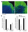
Homeostatic chemokine suppression is dependent on TLR4 expression. (a) Tissue sections from TLR4-KO mice before and 24 h after bacterial instillation, stained to reveal DLN architecture. (B cell zones: B220, green; T cell zones: CD3, blue.) Scale bar, 200 µm. (b) Relative mRNA levels in WT or TLR4-KO mice infected with S. typhimurium versus saline-injected controls. *P < 0.05; error bars represent means ± s.e.m. Similar data were acquired in three independent trials.
Figure 4
Figure 4
Socs3 and Smad3 modulate LPS-mediated CCL21 suppression. (a) DLN sections, 24 h after footpad injection of 1 × 10⁵ CFU of S. typhimurium, stained for the HEV marker PNAd and Socs3. The bottom panels show co-localization of these markers by multi-channel image (left) and software-generated co-localization image (right). Scale bar, 25 nm. (b) Expression of Socs3 in SVEC4-10 cells 6 h after treatment with WT or ΔmsbB S. typhimurium or LPS. *P < 0.01. (c) Socs3 expression 6 h after treatment with (+) or without (−) sLPS (left) or CCL21 expression 24 h after treatment (right) in SVEC4-10 cells treated with siRNA. For left graph, *P < 0.001. For right graph, *P < 0.01. (d) Expression of Socs3 (left) and CCL21 (right) in Socs3-overexpressing (Socs3-O) cells compared to control cells. *P = 0.02. (e) Western blots, probed using an antibody against P-Smad3, after SDS-PAGE separation of proteins from SVEC4-10 cells treated with WT or ΔmsbB S. typhimurium or sLPS. Blots contain either cytoplasmic fractions or proteins pulled down from nuclear fractions using an antibody against Smad3. (f) Relative Smad3 and CCL21 expression, determined by real-time PCR, after Smad3 knockdown (KD) in SVEC4-10 cells using siRNA. *P = 0.002. (g) Relative levels of Smad3 and CCL21 in SVEC4-10 cells overexpressing Smad3 (Smad-O). For Smad3 expression, *P = 0.03. For CCL21 expression, *P = 0.04. For all panels, error bars represent means ± s.e.m., and data are representative of three or more trials.
Figure 5
Figure 5
Adaptive immune contributions to limiting S. typhimurium infection. (a) Survival of mice inoculated orally with 1 × 10⁴ CFU of WT S. typhimurium compared to mice challenged with the ΔmsbB strain. (b) Quantification of bacterial numbers in the liver and spleen after 4 d of infection by WT or ΔmsbB S. typhimurium. (c) Quantification of bacterial numbers in the spleen and liver in mice infected with ΔmsbB S. typhimurium that had undergone combined or individual chemokine blockade or control antibody (Ig) administration (see Online Methods). P < 0.03, by one-way analysis of variance. See Supplementary Table 1 for further statistical analysis of panels b and c. For b and c, error bars represent means ± s.e.m., and data are representative of two or more independent experiments, except in the case individual chemokine blockade, included in one trial for a total n = 3. (d) Survival curves of mice treated with control or combined chemokine-specific antibody treatment, P = 0.02. (e) Survival of nude mice on a C57BL/6J background compared to heterozygous control mice after oral infection with the ΔmsbB strain. P = 0.05. (f) Survival of nude mice after oral infection with the ΔmsbB strain compared to WT mice infected orally with WT S. typhimurium. P = 0.47.
Figure 6
Figure 6
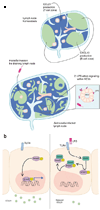
Model of S. typhimurium-induced suppression of homeostatic chemokines. (a) Schematic illustrating the proposed mechanism of S. typhimurium–induced lymph node architecture changes. During lymph node homeostasis, the expression of chemokines within the DLN contributes to the maintenance of normal architecture. CCL21 and CXCL13 are two key chemokines that promote the formation of T and B cell zones, respectively. Subsequent to the initial invasion of S. typhimurium into the host peripheral tissues, S. typhimurium invades further into the host, reaching the DLN. Once in the DLN, S. typhimurium’s presence disrupts cellular trafficking within the DLN, characterized by disorganized B and T cell zones and altered DLN cellular constituents. The disruption of S. typhimurium–infected DLNs is accompanied by a reduced expression of CCL21 and CXCL13. Our data suggests that the mechanism of CCL21 reduction within HEVs is dependent on LPS signaling through TLR4. (b) In the depicted model of TLR4 signaling, engagement of TLR4 results in upregulation of Socs3, a negative regulator of the transcription factor, Smad3. At basal levels, phosphorylated Smad3 is able to enter the nucleus, where it functions upstream of CCL21 expression in the SVEC4-10 model of HEVs. The upregulation of Socs3 in response to TLR4 signaling results in sequestration of Smad3 in the cytoplasm and reduces Smad3 promoted CCL21 expression.

References

    1. Gunn MD, et al. A chemokine expressed in lymphoid high endothelial venules promotes the adhesion and chemotaxis of naive T lymphocytes. Proc. Natl. Acad. Sci. USA. 1998;95:258–263. - PMC - PubMed
    1. Gunn MD, et al. A B cell–homing chemokine made in lymphoid follicles activates Burkitt’s lymphoma receptor-1. Nature. 1998;391:799–803. - PubMed
    1. Cyster JG. Chemokines and cell migration in secondary lymphoid organs. Science. 1999;286:2098–2102. - PubMed
    1. von Andrian UH, Mempel TR. Homing and cellular traffic in lymph nodes. Nat. Rev. Immunol. 2003;3:867–878. - PubMed
    1. Cyster JG, et al. Follicular stromal cells and lymphocyte homing to follicles. Immunol. Rev. 2000;176:181–193. - PubMed

Publication types

MeSH terms

LinkOut - more resources