. 2009 Dec 17;28(50):4469-79.
doi: 10.1038/onc.2009.270. Epub 2009 Oct 26.

Silencing of the Lats2 tumor suppressor overrides a p53-dependent oncogenic stress checkpoint and enables mutant H-Ras-driven cell transformation

Affiliations

Silencing of the Lats2 tumor suppressor overrides a p53-dependent oncogenic stress checkpoint and enables mutant H-Ras-driven cell transformation

Y Aylon et al. Oncogene. .

Abstract

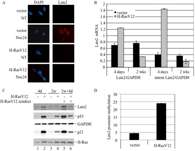
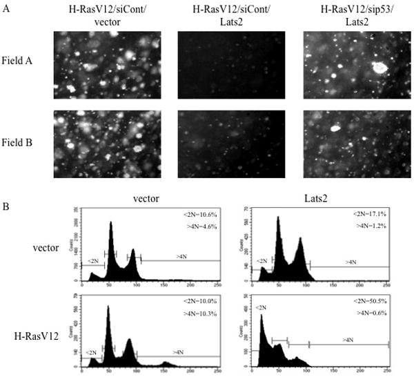
The Lats2 tumor suppressor protein has been implicated earlier in promoting p53 activation in response to mitotic apparatus stress, by preventing Mdm2-driven p53 degradation. We now report that Lats2 also has a role in an ATR-Chk1-mediated stress check point in response to oncogenic H-Ras. Activated mutant H-Ras triggers the translocation of Lats2 from centrosomes into the nucleus, coupled with an increase in Lats2 protein levels. This leads to the induction of p53 activity, upregulation of proapoptotic genes, downregulation of antiapoptotic genes and eventually apoptotic cell death. Many of the cells that survive apoptosis undergo senescence. However, a fraction of the cells escape this checkpoint mechanism, despite maintaining a high mutant H-Ras expression. These escapers display increased genome instability, as evidenced by a substantial fraction of cells with micronuclei and cells with polyploid genomes. Interestingly, such cells show markedly reduced levels of Lats2, in conjunction with enhanced hypermethylation of the Lats2 gene promoter. Our findings suggest that Lats2 might have an important role in quenching H-Ras-induced transformation, whereas silencing of Lats2 expression might serve as a mechanism to enable tumor progression.


Figures

Figure 1
Figure 1. H-Ras overexpression causes an increase in endogenous Lats2
(A) WI-38 cells were infected with H-RasV12 or vector only. Lysates of hygromycin-resistant cells were analyzed four days after infection by Western blot to visualize Lats2 protein, GAPDH and H-Ras. (B) Cells were infected as in (A). RNA was prepared from each culture four days after infection and analyzed by qRT-PCR. Values were normalized to GAPDH mRNA. “Lats2” refers to the product of primers amplifying the exon3-exon4 junction whereas “intron Lats2” amplifies a region within intron 3 of Lats2. (C) Cells were infected as in (A), treated four days after infection with 80μg/ml cycloheximide (CHX) for the indicated time periods, and then harvested for Western blot analysis. For easier comparison, both a short and long exposure of the Lats2 blot are presented.
Figure 2
Figure 2. Chk1 and ATR are implicated in Lats2 accumulation in response to activated H-Ras
(A) Cells were infected as in Fig. 1A. Four days after infection, hygromycin-resistant cultures were treated overnight with either 10μM SB 203580, 0.5μM UCN-01, or 100μM caffeine, or left non-treated (mock). Cell lysates were subjected to Western blot analysis to visualize ATR, phospho-ATR, total Chk1, phospho-Chk1, p38 and phopho-p38. (B) Cells were processed and treated as in (A), fixed and immunostained to visualize Lats2. Nuclear DNA was visualized by DAPI staining. Arrowhead denotes cell with micronuclei. Protein signals were quantified using ImageJ software and normalized to the corresponding GAPDH signals.
Figure 3
Figure 3. p53 response to H-RasV12 overexpression is dependent on Lats2
(A) WI-38 cells were infected as in Fig. 1A. Four days after infection, hygromycin-resistant cells were transiently transfected with Lats2 siRNA or control siRNA. Cell lysates were prepared 24 hours after transfection and analyzed by Western blot to visualize Lats2, p53 and GAPDH. (B) Cells were processed as in (A) and subjected to Western blot analysis with antibodies against Lats2, β-tubulin, p21 and H-Ras. (C) Cells were processed as in (A). RNA was prepared 24 hours after transfection and subjected to qRT-PCR analysis with primers specific for the indicated genes. Values were normalized to HPRT mRNA. (D) Cells were processed as in (A), except that transfection was with either LacZ siRNA, Lats2 siRNA or p53 siRNA. RNA was prepared 24 hours after transfection and subjected to qRT-PCR analysis with primers specific for the indicated genes. Values were normalized to HPRT mRNA.
Figure 4
Figure 4. Lats2 affects the biological outcome of H-RasV12 overexpression
(A) WI-38 cells were infected with various combinations of recombinant retroviruses encoding H-RasV12, Lats2 shRNA (siLats2) or mouse-specific PUMA shRNA (siCont) or empty retrovirus (vector). Cells were fixed, stained with PI and taken for FACS analysis either 4 days after infection and hygromycin and blasticidin selection or after an additional two weeks (2 wks) of growth in culture with antibiotic selection. Positions and percentages of sub-G1 cells (<2N) and polyploid cells (>4N) are indicated. Data is representative of at least three independent experiments. (B) Cells were infected with empty retrovirus (vector) or H-RasV12 and selected with hygromycin. Four days after infection, cultures were treated for 24 hours with 100μM caffeine or left untreated. Cells were fixed, stained with PI and subjected to FACS analysis. (C) Cells were infected and selected as in (B). Four days after infection, cells were transiently transfected with Lats2 siRNA or lacZ siRNA. 24 hours after transfection equal amounts of cells were plated in transwells and subjected in duplicate to migration assays. Wells were photographed at two magnifications as indicated. Data is representative of at least three independent experiments. (D) Cells were infected and processed as in (A). Cultures were stained for β-gal activity after 10 days of combined hygromycin and blasticidin selection. (E) Cells were infected and selected as in (D). Data represents percentage of β-gal positive cells after 4 and 10 days of H-RasV12 expression, based on counting of blue and white cells from three randomly chosen fields from each condition. Error bars represent standard deviation.
Figure 5
Figure 5. Chronic expression of H-RasV12 selects for Lats2 silencing
(A) WI-38 cells were infected with either vector only or H-RasV12. After three weeks of selection, cells were treated overnight with 200ng/ml nocodazole (NOC) or DMSO only (NT). Fixed cells were immunostained to visualize Lats2. Nuclear DNA was visualized by DAPI staining. (B) Cells were infected as in (A). RNA was analyzed by qRT-PCR following 4 days or 2 weeks of selection, using exon-junction Lats2 primers (left) or intronic Lats2 primers (right). In both instances, values were normalized to GAPDH mRNA. (C) Cells were infected as in (A). After two weeks of growth under hygromycin selection, cells were infected with a recombinant retrovirus expressing H-RasV12 carrying puromycin resistance and grown for an additional 4 days (2w+4d). Cell lysates were subjected to Western blot analysis to visualize Lats2, p53, GAPDH, p21 and H-Ras. (D) Cells were infected as in (A). After two weeks of selection, high molecular weight DNA was extracted, treated with bisulfate and analyzed by qRT-PCR using methylation-specific primers. Values were normalized to non-methylated ALU sequence and a total-methylated SssI sample.
Figure 6
Figure 6. Overexpression of Lats2 prevents soft agar colony formation in a p53-dependent manner and triggers apoptosis
(A) WI-38 cells were infected with various combinations of recombinant retroviruses encoding H-RasV12, Myc-tagged Lats2 (Lats2), p53 shRNA (sip53), mouse-specific PUMA shRNA (siCont) or empty retrovirus (vector). Following selection for two weeks under hygromycin and blasticidin, equal amounts of cells were subjected in duplicate to a soft agar colony formation assay. Four weeks after plating, microcolonies were photographed. Two representative fields are shown for each condition. (B) WI-38 cells were infected with vector only or H-RasV12. Following three weeks of hygromycin selection, cells were infected with vector only or Myc-tagged Lats2 (Lats2) and subjected to blasticidin selection. Cells were harvested 4 days after the second infection, fixed, stained with PI and subjected to FACS analysis.

References

    1. Abulaiti A, Fikaris AJ, Tsygankova OM, Meinkoth JL. Ras induces chromosome instability and abrogation of the DNA damage response. Cancer Res. 2006;66(21):10505–10512. - PubMed
    1. Aylon Y, Michael D, Shmueli A, Yabuta N, Nojima H, Oren M. A positive feedback loop between the p53 and Lats2 tumor suppressors prevents tetraploidization. Genes Dev. 2006;20(19):2687–2700. - PMC - PubMed
    1. Bartek J, Bartkova J, Lukas J. DNA damage signalling guards against activated oncogenes and tumour progression. Oncogene. 2007;26(56):7773–7779. - PubMed
    1. Bartkova J, Rezaei N, Liontos M, Karakaidos P, Kletsas D, Issaeva N, et al. Oncogene-induced senescence is part of the tumorigenesis barrier imposed by DNA damage checkpoints. Nature. 2006;444(7119):633–637. - PubMed
    1. Bean GR, Bryson AD, Pilie PG, Goldenberg V, Baker JC, Jr, Ibarra C, et al. Morphologically normal-appearing mammary epithelial cells obtained from high-risk women exhibit methylation silencing of INK4a/ARF. Clin Cancer Res. 2007;13(22 Pt 1):6834–6841. - PubMed

Publication types

MeSH terms