. 2010 Jan 21;29(3):335-44.
doi: 10.1038/onc.2009.333. Epub 2009 Oct 26.

Activated BRAF induces gliomas in mice when combined with Ink4a/Arf loss or Akt activation

Affiliations

Activated BRAF induces gliomas in mice when combined with Ink4a/Arf loss or Akt activation

J P Robinson et al. Oncogene. .

Abstract

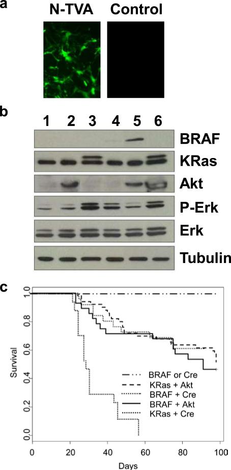
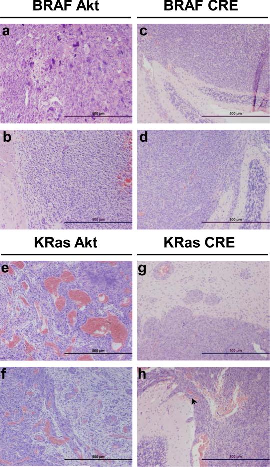
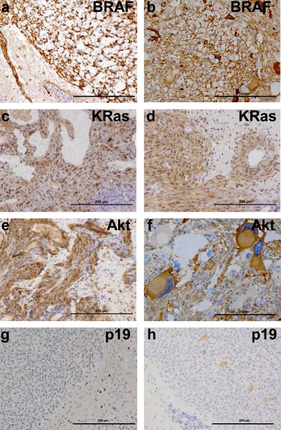
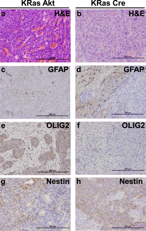
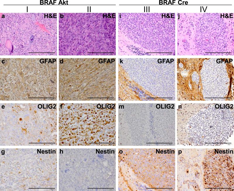
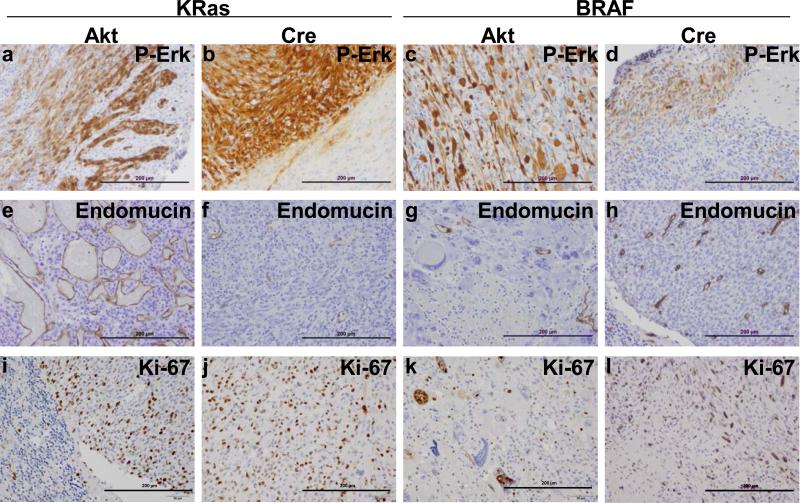
Mutations in receptor tyrosine kinase (RTK) growth factor receptors (epidermal growth factor receptor, platelet-derived growth factor receptor, MET and ERBB2), which result in downstream activation of the RAS/RAF/MEK/ERK mitogen-activated protein kinase (MAPK) pathway and PI(3)K/Akt pathway, are found in almost all high-grade gliomas and MAPK signaling is necessary for continued glioma maintenance. In addition, BRAF is mutated in the majority of low-grade gliomas and its expression and activity is significantly increased in the majority of high-grade gliomas. Although the importance of RTKs and RAS signaling in glioma development has been shown, the role of BRAF has yet to be characterized. We evaluated the effect of activated BRAF in glioma formation using the retroviral replication-competent avian leukosis virus long terminal repeat, splice acceptor (RCAS)/TVA system to transfer genes encoding activated forms of BRAF, KRas, Akt and Cre to nestin-expressing neural progenitor cells in Ink4a/Arf(lox/lox) mice in vivo. Although expression of activated BRAF alone is not sufficient for tumorigenesis, the combination of activated BRAF and Akt or BRAF with Ink4a/Arf loss is transforming. Interestingly, activated BRAF generates gliomas with characteristics similar to activated KRas in the context of Akt but not Ink4a/Arf loss. Our studies show a role for BRAF activation and signaling in glioma development and as potential target for glioma therapy.


Figures

Figure 1
Figure 1. KRasG12D and BRAFV600E cooperate with activated Akt or Ink4a/Arf loss to induce gliomas in N-TVA mice
(a) In vitro expression of GFP in N-TVA astrocytes. RCASBP(A)GFP infection of N-TVA positive and negative (control) cells in culture confirmed stable and targeted gene delivery of GFP into N-TVA expressing cells. (b) Western blot analysis of gene expression. N-TVA-positive immortalized astrocytes were infected in culture with RCASBP(A)GFP (lane 1), RCASBP(A)Akt (lane 2), RCASBP(A)KRasG12D (lane 3), RCASBP(A)BRAFV600E (lane 4), RCASBP(A)BRAFV600E and RCASBP(A)Akt (lane 5), or RCASBP(A)Akt and RCASBP(A)KRasG12D (lane 6). Lysates were separated on a 4-20% Tris-glycine polyacrylamide gel, transferred to nitrocellulose, and probed with antibodies directed against V5 (BRAF), HA (Akt), pan-RAS (KRas), phoshporylated Erk (P-Erk), or total Erk (Erk) as indicated. The membrane was re-probed with an antibody against α-tubulin (bottom) to ensure equal loading. The Ras blot shows endogenous Ras (lower band) and the virally delivered KRas (upper band). The virally delivered KRas is a higher molecular weight as a result of the FLAG epitope tag. (c) Kaplan-Meier percent survival curve. N-TVA;Ink4a/Arflox/lox mice were injected with the indicated viruses at birth. Censored survival data was analyzed using a log-rank test of the Kaplan-Meier estimate of survival. No tumors were observed in mice injected with BRAFV600E or Cre alone n = 36 (solid black line) and it has previously been shown that no tumors form in mice injected with either KRasG12D or Akt alone (Holland et al., 2000). There was no significant difference in survival between KRasG12D + Akt n = 50 (red line) BRAFV600E + Cre n = 26 (blue line) or BRAFV600E + Akt n = 28 (green line). A significant difference in survival was observed between KRasG12D + Cre n = 17 (dashed black line) and all of the other conditions, P < 3 × 10⁻⁷.
Figure 2
Figure 2. Representative tumors induced by KRasG12D or BRAFV600E in cooperation with activated Akt or Cre (Ink4a/Arf loss)
Histological comparison of gliomas induced in N-TVA;Ink4a/Arflox/lox mice after injection with (a-b) BRAFV600E + Akt, (c-d) BRAFV600E + Cre, (e-f) KRasG12D + Akt and (g-h) KRasG12D + Cre. Although the CNS border was well defined (g-h) and the tumors were not infiltrative, invasion was seen to progress through the subarachnoid space and Virchow-Robin perivascular spaces (arrow, h). Scale bars represent 500 μm.
Figure 3
Figure 3. Immunohistochemical confirmation of gene expression in gliomas arising in N-TVA;Ink4a/Arflox/lox mice
(a) IHC for the V5 epitope tag on BRAFV600E in gliomas induced by BRAFV600E + Cre. (b) IHC for the V5 epitope tag on BRAFV600E in gliomas induced by BRAFV600E + Akt. (c) IHC for the FLAG epitope tag on KRas in gliomas induced by KRasG12D +Akt. (d) IHC for the FLAG epitope tag on KRasG12D in gliomas induced by KRasG12D + Cre. (e) IHC for the HA epitope tag on Akt in gliomas induced by KRasG12D + Akt. (f) IHC for the HA epitope tag on Akt in gliomas induced by BRAFV600E + AKT. IHC for p19 indicates loss of p19 expression in (g) KRasG12D, (f) BRAFV600E + Cre induced gliomas. Scale bars represent 200 μm.
Figure 4
Figure 4. Expression of astrocyte and oligodendrocyte markers in gliomas induced with KRasG12D
(a-b) Corresponding H&E sections for histological comparison with IHC analysis. (c-d) Expression of the astrocyte marker GFAP. (e-f) Expression of the oligodendrocyte marker Olig2. (g-h) Expression of the neural progenitor cell marker Nestin. Scale bars represent 200 μm.
Figure 5
Figure 5. Expression of astrocyte and oligodendrocyte markers in gliomas induced with BRAFV600E
(I-II) BRAFV600E gliomas induced with activated Akt were highly pleomorphic in expression and appearance so two examples are highlighted for comparison with (III-IV) the highly monomorphic nature of BRAFV600E gliomas in the context of Ink4a/Arf loss. (a-b and i-j) Corresponding H&E sections for histological comparison with IHC analysis. (c-f and k-l) Expression of the astrocyte marker GFAP. (e-f and m-n) Expression of the oligodendrocyte marker Olig2. (g-h and o-p) Expression of the neural progenitor cell marker Nestin. Scale bars represent 200 μm.
Figure 6
Figure 6. All gliomas demonstrate MAPK pathway activation and high levels of proliferation
(A-D) Confirmation of MAPK activation. IHC for P-Erk confirmed MAPK activation in all gliomas induced with either KRasG12D or BRAFV600E. (e-h) Analysis of tumor vasculature. IHC for the endothelial cell marker endomucin highlights the highly abnormal vasculature of Akt/KRasG12D gliomas compared with the other tumor samples. (i-l) Assessment of mitotic activity. IHC for the cell division marker Ki67 demonstrates that all of the tumors are highly proliferative. Scale bars represent 200 μm.

References

    1. Aguirre AJ, Bardeesy N, Sinha M, Lopez L, Tuveson DA, Horner J, et al. Activated Kras and Ink4a/Arf deficiency cooperate to produce metastatic pancreatic ductal adenocarcinoma. Genes Dev. 2003;17:3112–26. - PMC - PubMed
    1. Balmanno K, Cook SJ. Tumour cell survival signalling by the ERK1/2 pathway. Cell Death Differ. 2009;16:368–77. - PubMed
    1. Barnier JV, Papin C, Eychene A, Lecoq O, Calothy G. The mouse B-raf gene encodes multiple protein isoforms with tissue-specific expression. J Biol Chem. 1995;270:23381–9. - PubMed
    1. Basto D, Trovisco V, Lopes JM, Martins A, Pardal F, Soares P, et al. Mutation analysis of B-RAF gene in human gliomas. Acta Neuropathol. 2005;109:207–10. - PubMed
    1. Bromberg-White JL, Webb CP, Patacsil VS, Miranti CK, Williams BO, Holmen SL. Delivery of short hairpin RNA sequences by using a replication-competent avian retroviral vector. J Virol. 2004;78:4914–6. - PMC - PubMed

Publication types

MeSH terms

Substances