

Sulf-2, a heparan sulfate endosulfatase, promotes human lung carcinogenesis
- PMID: 19855436
- PMCID: PMC2818095
- DOI: 10.1038/onc.2009.365
Sulf-2, a heparan sulfate endosulfatase, promotes human lung carcinogenesis
Abstract
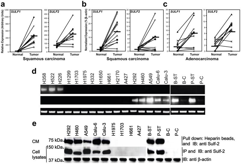
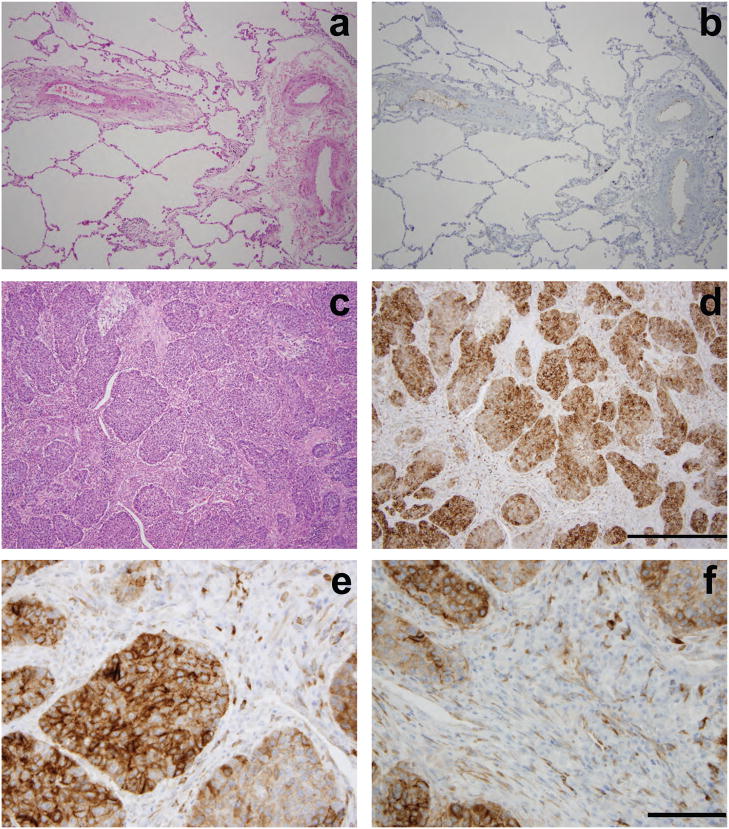
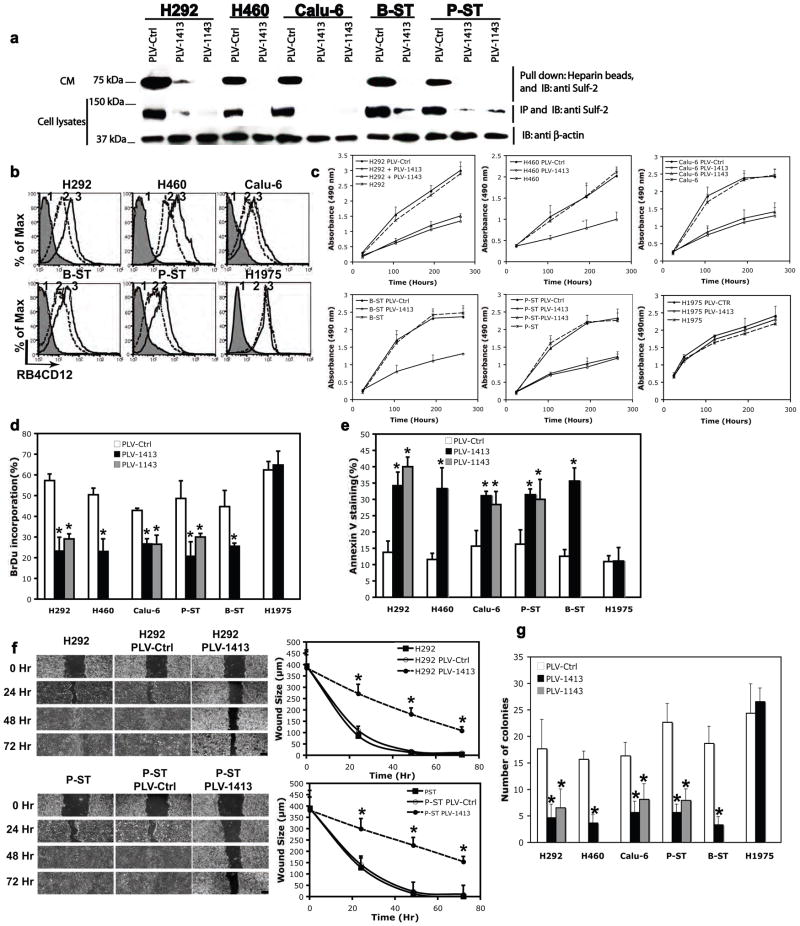
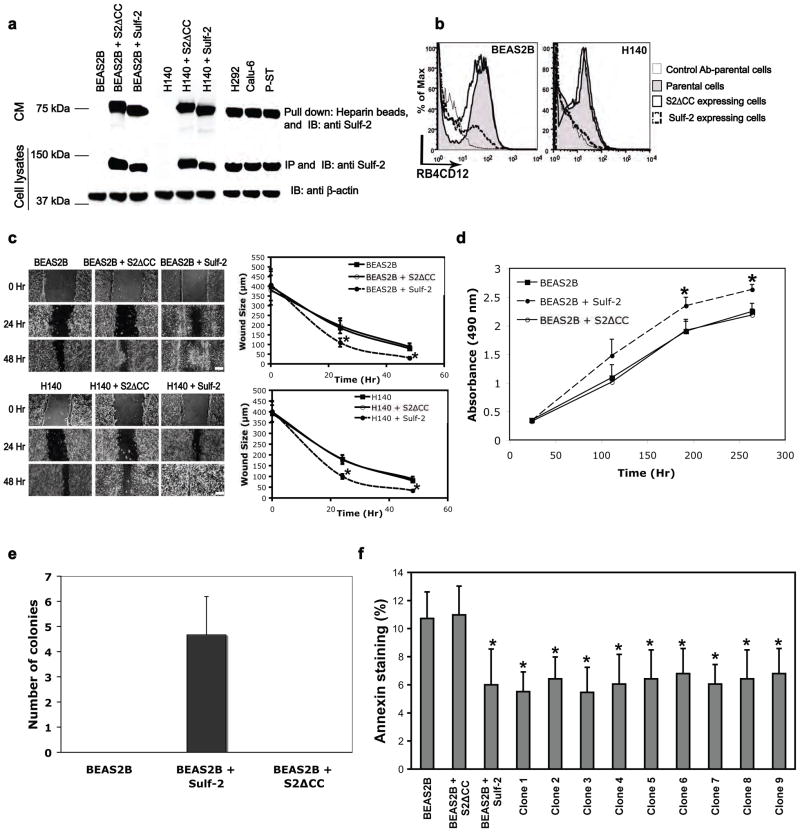
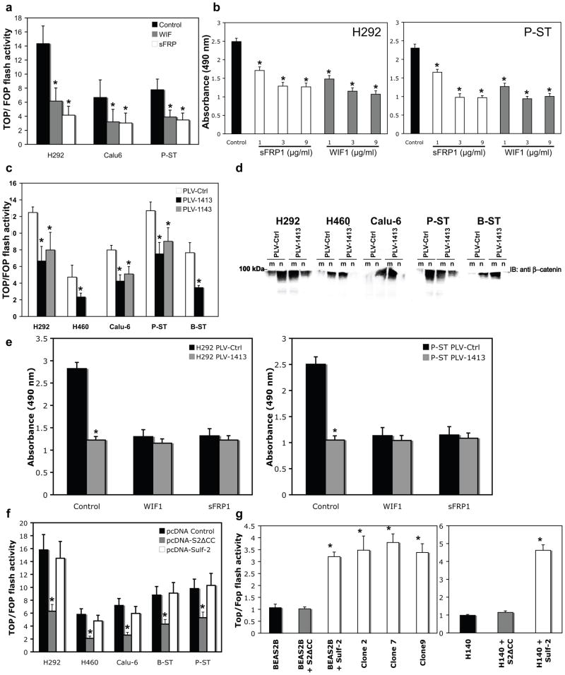
Heparan sulfate (HS) proteoglycans (HSPGs) bind to multiple growth factors/morphogens and regulate their signaling. 6-O-sulfation (6S) of glucosamine within HS chains is critical for many of these ligand interactions. Sulf-1 and Sulf-2, which are extracellular neutral-pH sulfatases, provide a novel post-synthetic mechanism for regulation of HSPG function by removing 6S from intact HS chains. The Sulfs can thereby modulate several signaling pathways, including the promotion of Wnt signaling. We found induction of SULF2 transcripts and Sulf-2 protein in human lung adenocarcinoma and squamous cell carcinoma, the two major classes of non-small-cell lung carcinomas (NSCLCs). We confirmed widespread Sulf-2 protein expression in tumor cells of 10/10 surgical specimens of human lung squamous carcinomas. We studied five Sulf-2(+) NSCLC cell lines, including two, which were derived by cigarette-smoke transformation of bronchial epithelial cells. shRNA-mediated Sulf-2 knockdown in these lines caused an increase in 6S on their cell surface and in parallel reversed their transformed phenotype in vitro, eliminated autocrine Wnt signaling and strongly blunted xenograft tumor formation in nude mice. Conversely, forced Sulf-2 expression in non-malignant bronchial epithelial cells produced a partially transformed phenotype. Our findings support an essential role for Sulf-2 in lung cancer, the leading cancer killer.
Figures

Similar articles
-
Sulf loss influences N-, 2-O-, and 6-O-sulfation of multiple heparan sulfate proteoglycans and modulates fibroblast growth factor signaling.J Biol Chem. 2008 Oct 10;283(41):27724-27735. doi: 10.1074/jbc.M802130200. Epub 2008 Aug 6. J Biol Chem. 2008. PMID: 18687675
-
The SULFs, extracellular sulfatases for heparan sulfate, promote the migration of corneal epithelial cells during wound repair.PLoS One. 2013 Aug 8;8(8):e69642. doi: 10.1371/journal.pone.0069642. eCollection 2013. PLoS One. 2013. PMID: 23950901 Free PMC article.
-
Extracellular sulfatases, elements of the Wnt signaling pathway, positively regulate growth and tumorigenicity of human pancreatic cancer cells.PLoS One. 2007 Apr 25;2(4):e392. doi: 10.1371/journal.pone.0000392. PLoS One. 2007. PMID: 17460759 Free PMC article.
-
Sulf-2: an extracellular modulator of cell signaling and a cancer target candidate.Expert Opin Ther Targets. 2010 Sep;14(9):935-49. doi: 10.1517/14728222.2010.504718. Expert Opin Ther Targets. 2010. PMID: 20629619 Free PMC article. Review.
-
Use of a phage display antibody to measure the enzymatic activity of the Sulfs.Methods Enzymol. 2010;480:51-64. doi: 10.1016/S0076-6879(10)80003-5. Methods Enzymol. 2010. PMID: 20816204 Review.
Cited by
-
The prognostic value and immunological role of SULF2 in adrenocortical carcinoma.Heliyon. 2023 Feb 11;9(2):e13613. doi: 10.1016/j.heliyon.2023.e13613. eCollection 2023 Feb. Heliyon. 2023. PMID: 36852051 Free PMC article.
-
Changes in heparan sulfate sulfotransferases and cell-surface heparan sulfate during SKM-1 cells granulocytic differentiation and A549 cells epithelial-mesenchymal transition.Glycoconj J. 2020 Apr;37(2):151-164. doi: 10.1007/s10719-019-09903-0. Epub 2019 Dec 20. Glycoconj J. 2020. PMID: 31863309
-
The oncogenic effect of sulfatase 2 in human hepatocellular carcinoma is mediated in part by glypican 3-dependent Wnt activation.Hepatology. 2010 Nov;52(5):1680-9. doi: 10.1002/hep.23848. Hepatology. 2010. PMID: 20725905 Free PMC article.
-
Post-Synthetic Regulation of HS Structure: The Yin and Yang of the Sulfs in Cancer.Front Oncol. 2014 Jan 14;3:331. doi: 10.3389/fonc.2013.00331. eCollection 2014 Jan 14. Front Oncol. 2014. PMID: 24459635 Free PMC article. Review.
-
SULF2 methylation is prognostic for lung cancer survival and increases sensitivity to topoisomerase-I inhibitors via induction of ISG15.Oncogene. 2012 Sep 13;31(37):4107-16. doi: 10.1038/onc.2011.577. Epub 2011 Dec 12. Oncogene. 2012. PMID: 22158045 Free PMC article.
References
-
- Ai X, Kitazawa T, Do A-T, Kusche-Gullberg M, Labosky PA, Emerson CP., Jr SULF1 and SULF2 regulate heparan sulfate-mediated GDNF signaling for esophageal innervation. Development. 2007;134:3327–3338. - PubMed
-
- Alexander CM, Reichsman F, Hinkes MT, Lincecum J, Becker KA, Cumberledge S, et al. Syndecan-1 is required for Wnt-1-induced mammary tumorigenesis in mice. Nat Genet. 2000;25:329–332. - PubMed
-
- Bafico A, Liu G, Goldin L, Harris V, Aaronson SA. An autocrine mechanism for constitutive Wnt pathway activation in human cancer cells. Cancer Cell. 2004;6:497–506. - PubMed
Publication types
MeSH terms
Substances
Grants and funding
LinkOut - more resources
Full Text Sources
Other Literature Sources
Medical
