

Review
doi: 10.1007/s00467-009-1320-9.
Epub 2009 Oct 24.
Imaging in the evaluation of renovascular disease
Affiliations
- PMID: 19856000
- PMCID: PMC2855432
- DOI: 10.1007/s00467-009-1320-9
Item in Clipboard
Review
Imaging in the evaluation of renovascular disease
Pediatr Nephrol.
2010 Jun.
Abstract
Renovascular disease (RVD) is an important cause of hypertension in children, as it often is amenable to potentially curative treatment. Imaging aimed at finding RVD therefore needs to have high sensitivity so as not to miss important findings. Digital subtraction angiography is the gold standard investigation. Doppler ultrasonography, computed tomography (CT) angiography and magnetic resonance (MR) angiography can all be helpful, but none has, at present, high enough sensitivity to rule out RVD in a child with a suggestion of that diagnosis.
Figures
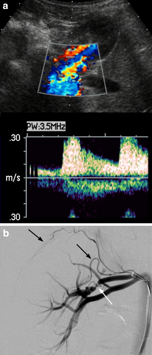
A 6-year-old boy with hypertension. a Doppler ultrasound of the right renal artery or one of its major branches shows a normal waveform. b Digital subtraction angiography shows a critical stenosis of a large branch of the right renal artery (white arrow), associated with tiny aneurysms. Collateral vessels are seen, both locally and arising from a capsular artery (black arrows)
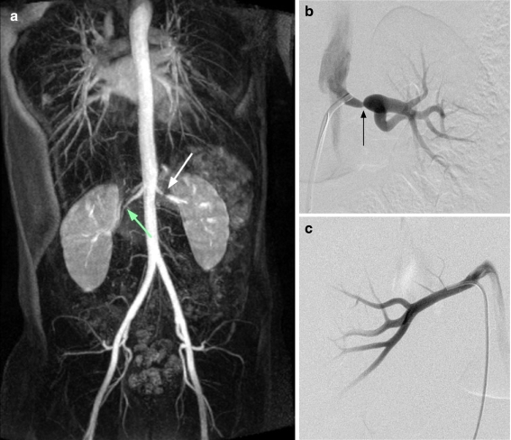
An 11-year-old boy with hypertension. a Contrast-enhanced magnetic resonance angiography (coronal maximum intensity projection image) shows stenosis of the left renal artery (white arrow) but also suggests stenosis of the right (green arrow). b Angiogram of left kidney confirms a tight stenosis of the left main renal artery (arrow). c The right renal artery is normal
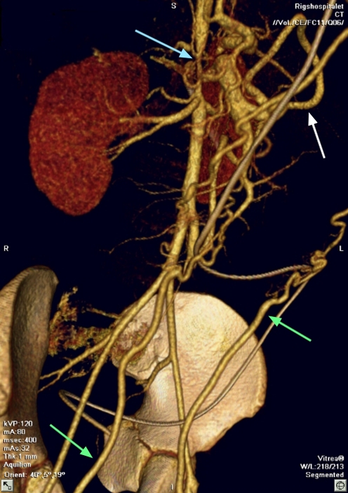
Two-dimensional (‘volume-rendered’) representation of a three-dimensional computed tomography dataset from a 12-year-old boy. There is severe stenosis of the abdominal aorta (‘mid-aortic syndrome’, blue arrow). Enlarged collateral arteries are present: the marginal artery of Drummond (white arrow) connects the superior and inferior mesenteric arteries, and the epigastric arteries (green arrows) connect the subclavian and femoral arteries on each side. Note that, although the coverage is outstanding, detail of the intra-renal arteries is lost
References
Publication types
MeSH terms
LinkOut - more resources
Full Text Sources
Other Literature Sources
Medical
