

Relationship of temporal resolution to diagnostic performance for dynamic contrast enhanced MRI of the breast
- PMID: 19856413
- PMCID: PMC2935260
- DOI: 10.1002/jmri.21947
Relationship of temporal resolution to diagnostic performance for dynamic contrast enhanced MRI of the breast
Abstract
Purpose: To investigate the relationship between temporal resolution of dynamic contrast-enhanced (DCE) magnetic resonance imaging (MRI) and classification of breast lesions as benign versus malignant.
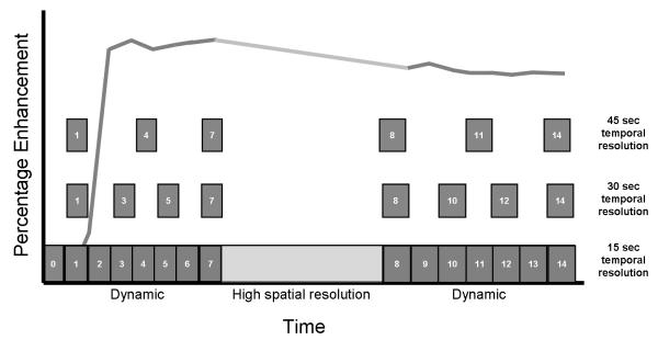
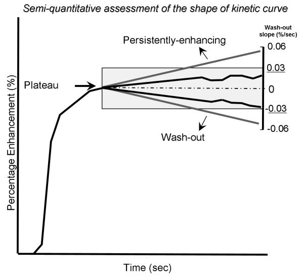
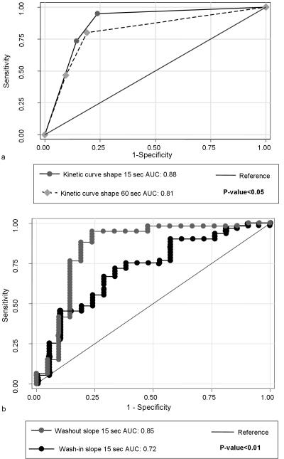
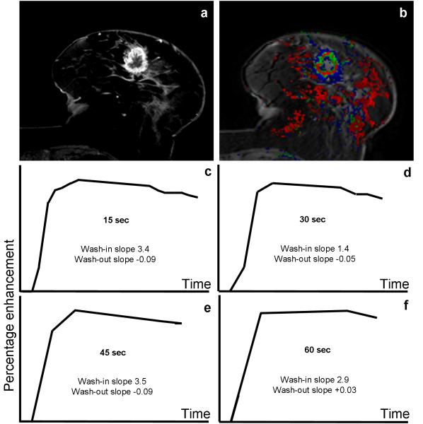
Materials and methods: Patients underwent T(1)-weighted DCE MRI with 15 s/acquisition temporal resolution using 1.5 Tesla (n = 48) and 3.0T (n = 33) MRI scanners. Seventy-nine patients had pathologically proven diagnosis and 2 had 2 years follow-up showing no change in lesion size. The temporal resolution of DCE MRI was systematically reduced as a postprocessing step from 15 to 30, 45, and 60 s/acquisition by eliminating intermediate time points. Average wash-in and wash-out slopes, wash-out percentage changes, and kinetic curve shape (persistently enhancing, plateau, or wash-out) were compared for each temporal resolution. Logistic regression and receiver operating characteristic (ROC) curve analysis were used to compare kinetic parameters and diagnostic accuracy.
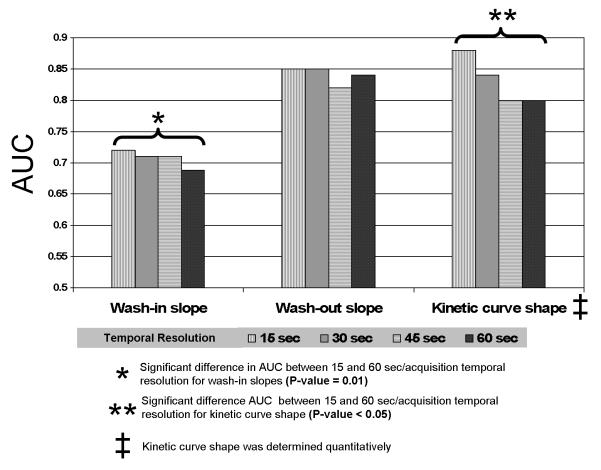
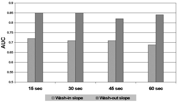
Results: Sixty patients (74%) had malignant lesions and 21 patients (26%) had benign lesions. All temporal-resolution parameters significantly predicted benign versus malignant diagnosis (P < 0.05). However, 45 s/acquisition and higher temporal-resolution datasets showed higher accuracy than the 60 s/acquisition dataset by ROC curve analysis (0.72 versus 0.69 for average wash-in slope; 0.85 versus 0.82, for average wash-out slope; and 0.88 versus 0.80 for kinetic curve shape assessment, for 45 s/acquisition versus 60 s/acquisition temporal-resolution datasets, respectively (P = 0.027).
Conclusion: DCE MRI data with at least 45-s temporal resolution maximized the agreement between the kinetic parameters and correct classification of benign versus malignant diagnosis.
Figures
References
-
- Sarrazin D, Le MG, Arriagada R, et al. Ten-year results of a randomized trial comparing a conservative treatment to mastectomy in early breast cancer. Radiother Oncol. 1989;14:177–184. - PubMed
-
- Huang W, Fisher PR, Dulaimy K, Tudorica LA, O’Hea B, Button TM. Detection of breast malignancy: diagnostic MR protocol for improved specificity. Radiology. 2004;232:585–591. - PubMed
-
- Viehweg P, Lampe D, Buchmann J, Heywang-Kobrunner SH. In situ and minimally invasive breast cancer: morphologic and kinetic features on contrast-enhanced MR imaging. Magma. 2000;11:129–137. - PubMed
-
- Heywang-Kobrunner SH, Bick U, Bradley WG, Jr., et al. International investigation of breast MRI: results of a multicentre study (11 sites) concerning diagnostic parameters for contrast-enhanced MRI based on 519 histopathologically correlated lesions. Eur Radiol. 2001;11:531–546. - PubMed
-
- Kutschker C, Allgayer B, Hauck W. [Evaluating the morphology of uncertain breast tumors using color coded Doppler ultrasound] Ultraschall Med. 1996;17:18–22. - PubMed
Publication types
MeSH terms
Substances
Grants and funding
LinkOut - more resources
Full Text Sources
Medical
