. 2010 Jan 15;395(2):390-407.
doi: 10.1016/j.jmb.2009.10.045. Epub 2009 Oct 24.

The crystal structure of the novobiocin biosynthetic enzyme NovP: the first representative structure for the TylF O-methyltransferase superfamily

Affiliations

The crystal structure of the novobiocin biosynthetic enzyme NovP: the first representative structure for the TylF O-methyltransferase superfamily

Inmaculada Gómez García et al. J Mol Biol. .

Abstract

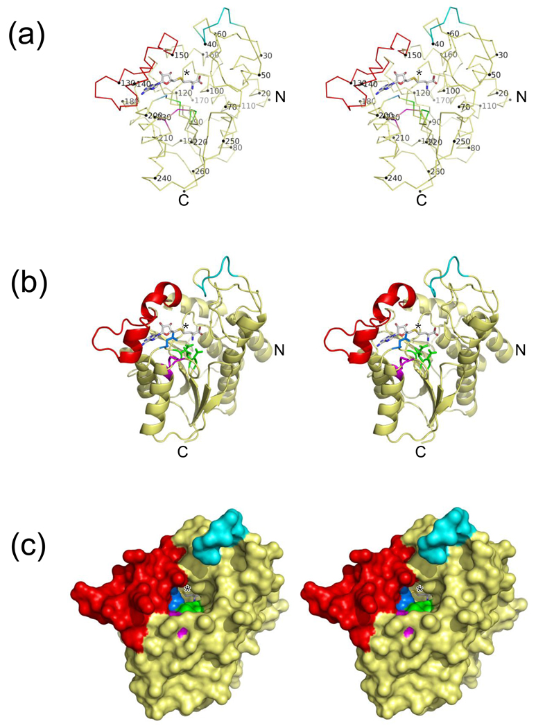
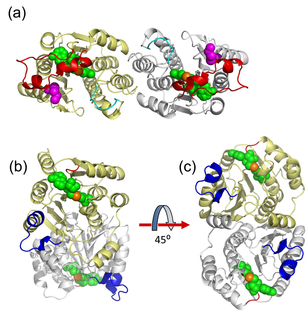
NovP is an S-adenosyl-l-methionine-dependent O-methyltransferase that catalyzes the penultimate step in the biosynthesis of the aminocoumarin antibiotic novobiocin. Specifically, it methylates at 4-OH of the noviose moiety, and the resultant methoxy group is important for the potency of the mature antibiotic: previous crystallographic studies have shown that this group interacts directly with the target enzyme DNA gyrase, which is a validated drug target. We have determined the high-resolution crystal structure of NovP from Streptomyces spheroides as a binary complex with its desmethylated cosubstrate S-adenosyl-l-homocysteine. The structure displays a typical class I methyltransferase fold, in addition to motifs that are consistent with a divalent-metal-dependent mechanism. This is the first representative structure of a methyltransferase from the TylF superfamily, which includes a number of enzymes implicated in the biosynthesis of antibiotics and other therapeutics. The NovP structure reveals a number of distinctive structural features that, based on sequence conservation, are likely to be characteristic of the superfamily. These include a helical 'lid' region that gates access to the cosubstrate binding pocket and an active center that contains a 3-Asp putative metal binding site. A further conserved Asp likely acts as the general base that initiates the reaction by deprotonating the 4-OH group of the noviose unit. Using in silico docking, we have generated models of the enzyme-substrate complex that are consistent with the proposed mechanism. Furthermore, these models suggest that NovP is unlikely to tolerate significant modifications at the noviose moiety, but could show increasing substrate promiscuity as a function of the distance of the modification from the methylation site. These observations could inform future attempts to utilize NovP for methylating a range of glycosylated compounds.


Figures

Fig. 1
Fig. 1
The structures of novobiocin and tylosin. (a) Novobiocin is an aminocoumarin antibiotic comprised of three ring systems, which are synthesised separately. The three methylation steps are indicated together with the final O-carbamoylation by NovN. NovO and NovU act during the synthesis of rings B and C, respectively. NovP methylates the 4-OH group of the L-noviose moiety when the antibiotic is almost fully formed; this represents the penultimate step of the whole pathway. The substrate for NovP, desmethyldescarbamoyl novobiocin (DDN) bears only hydroxyl groups at positions 3 and 4 of the noviose sugar. Rings B and C are identical in the substrates of the NovP orthologs CouP and CloP, with the exception that in the substrate for CloP, the C8 position of ring B is chlorinated rather than methylated. (b) Tylosin is a macrolide antibiotic. TylF performs the terminal step of the pathway by methylating the 3-OH group of 6-deoxy-2-O-methyl-D-allose.
Fig. 2
Fig. 2
Stereoviews showing the overall structure of NovP. (a) Backbone trace with every tenth residue labelled. (b) Cartoon representation with important residues shown in stick mode. (c) Molecular surface representation illustrating the solvent inaccessibility of the SAH and the shape of the active site channel. Throughout, the lid region is coloured red, and the disordered loop (residues 39–45 inclusive, which have been modelled into energetically favourable conformations) is shown in cyan. The 3-Asp putative metal-binding site is coloured green and Asp198, the proposed general base, is shown in blue. The disulphide bridge residues, Cys228 and Cys231, are coloured magenta. The SAH ligand is shown in stick representation and the position of the sulphur atom is indicated by an asterisk.
Fig. 3
Fig. 3
Multiple protein sequence alignments versus NovP. For both panels the alignment is displayed using ESPript. Strictly conserved residues are highlighted with red shaded boxes, and well conserved residues are boxed with the predominant residues coloured red. Secondary structure elements for NovP are shown above the alignment, where α = α helix, β = β strand, η = 310 helix, TT = β turn. Residues that are postulated to be involved in cation binding in NovP are marked by green triangles; Asp198, which is thought to act as a general base during the methyltransfer reaction, is indicated by the single blue triangle. Residues that are hydrogen bonded to SAH are indicated by dark blue letters where “S” refers to a side-chain interaction and “M” refers to a main-chain interaction. Although Asp196 is marked by a green triangle, indicating a role in cation binding, it also hydrogen bonds to SAH. Residues that form the NovP dimer interface are marked with asterisks, and these are coloured red for those residues that form hydrogen bonds. Other features of the NovP structure are labelled, i.e. the lid region, regions of disorder, the disulphide bridge, and the SAM-dependent methyltransferase motifs I and II. (a) Alignment of NovP with selected sequences from the TylF superfamily. The first four are from Streptomycetes (phylum Actinobacteria). They are: NovP from S. spheroides (NovP_S.sph), CouP from S. rishiriensis (CouP_S.ris), CloP from S, roseochromogenes subsp. oscitans (CloP_S.ros), and TylF from S. fradiae (TylF_S.fra). The remainder are representative sequences from the other six phyla which constitute the TylF superfamily, where the prefix "Hypo" indicates an uncharacterised or hypothetical protein. They are from: Bacillus anthracis (Hypo_B.ant; phylum Firmicutes), Herpetosiphon aurantiacus ATCC 23779 (Hypo_H.aur; phylum Chloroflexi), Trypanosoma cruzi (Hypo_T.cru; phylum Euglenozoa), Synechococcus sp. (strain JA-3-3Ab) (Hypo_S.sp.; phylum cyanobacteria), Rhodospirillum rubrum (Hypo_R.rub; phylum Proteobacteria) and Microscilla marina ATCC 23134 (MtfB_M.Mar; phylum Bacteroidetes). The magenta letters below the alignment indicate residues that delineate the substrate access channel, where "N" refers to those adjacent to the expected position of the noviose sugar (as predicted by the docking simulations), "A" refers to those adjacent to the expected position of the aminocoumarin ring, and "B" refers to the single residue, Trp58, that lies adjacent to both moieties. (b) Structure-based multiple sequence alignment of Streptomyces spheroides NovP (NovP_S.sph) with selected O-methyltransferase sequences for which there are crystal structures. Shown are the sequences from rat (COMT_R.nor), Synechocystis sp. strain PCC 6803 (SynOMT_S.sp), Mesembryanthemum crystallinum (PFOMT_M.cry), Leptospira interrogans (LiOMT_L.int), Bacillus cereus (BcOMT2_B.cer) and Medicago sativa (CCoAOMT_M.sat). These are also summarised in Table 1. The initial alignment was generated using EXPRESSO and subsequently adjusted manually with reference to the superposed structures. Indicated below the alignment is the N-terminal region of poor structural alignment (which includes helix α1), and the insertion loop that occurs in some of the structural homologues, but not NovP itself. Note that, Asp198, the putative general base (blue triangle), appears to be well conserved in the structural homologues of NovP. However, in these structures the protein backbone directs the Asp side-chain away from the active site. Instead, the side-chain of the next residue, a Lys in all but NovP, points into the active site and functions as the base. This is indicated by the yellow triangle shown below the alignment. This difference in main-chain conformation may be a consequence of the single residue insertion at the N-terminus of helix α9 in NovP.
Fig. 3
Fig. 3
Multiple protein sequence alignments versus NovP. For both panels the alignment is displayed using ESPript. Strictly conserved residues are highlighted with red shaded boxes, and well conserved residues are boxed with the predominant residues coloured red. Secondary structure elements for NovP are shown above the alignment, where α = α helix, β = β strand, η = 310 helix, TT = β turn. Residues that are postulated to be involved in cation binding in NovP are marked by green triangles; Asp198, which is thought to act as a general base during the methyltransfer reaction, is indicated by the single blue triangle. Residues that are hydrogen bonded to SAH are indicated by dark blue letters where “S” refers to a side-chain interaction and “M” refers to a main-chain interaction. Although Asp196 is marked by a green triangle, indicating a role in cation binding, it also hydrogen bonds to SAH. Residues that form the NovP dimer interface are marked with asterisks, and these are coloured red for those residues that form hydrogen bonds. Other features of the NovP structure are labelled, i.e. the lid region, regions of disorder, the disulphide bridge, and the SAM-dependent methyltransferase motifs I and II. (a) Alignment of NovP with selected sequences from the TylF superfamily. The first four are from Streptomycetes (phylum Actinobacteria). They are: NovP from S. spheroides (NovP_S.sph), CouP from S. rishiriensis (CouP_S.ris), CloP from S, roseochromogenes subsp. oscitans (CloP_S.ros), and TylF from S. fradiae (TylF_S.fra). The remainder are representative sequences from the other six phyla which constitute the TylF superfamily, where the prefix "Hypo" indicates an uncharacterised or hypothetical protein. They are from: Bacillus anthracis (Hypo_B.ant; phylum Firmicutes), Herpetosiphon aurantiacus ATCC 23779 (Hypo_H.aur; phylum Chloroflexi), Trypanosoma cruzi (Hypo_T.cru; phylum Euglenozoa), Synechococcus sp. (strain JA-3-3Ab) (Hypo_S.sp.; phylum cyanobacteria), Rhodospirillum rubrum (Hypo_R.rub; phylum Proteobacteria) and Microscilla marina ATCC 23134 (MtfB_M.Mar; phylum Bacteroidetes). The magenta letters below the alignment indicate residues that delineate the substrate access channel, where "N" refers to those adjacent to the expected position of the noviose sugar (as predicted by the docking simulations), "A" refers to those adjacent to the expected position of the aminocoumarin ring, and "B" refers to the single residue, Trp58, that lies adjacent to both moieties. (b) Structure-based multiple sequence alignment of Streptomyces spheroides NovP (NovP_S.sph) with selected O-methyltransferase sequences for which there are crystal structures. Shown are the sequences from rat (COMT_R.nor), Synechocystis sp. strain PCC 6803 (SynOMT_S.sp), Mesembryanthemum crystallinum (PFOMT_M.cry), Leptospira interrogans (LiOMT_L.int), Bacillus cereus (BcOMT2_B.cer) and Medicago sativa (CCoAOMT_M.sat). These are also summarised in Table 1. The initial alignment was generated using EXPRESSO and subsequently adjusted manually with reference to the superposed structures. Indicated below the alignment is the N-terminal region of poor structural alignment (which includes helix α1), and the insertion loop that occurs in some of the structural homologues, but not NovP itself. Note that, Asp198, the putative general base (blue triangle), appears to be well conserved in the structural homologues of NovP. However, in these structures the protein backbone directs the Asp side-chain away from the active site. Instead, the side-chain of the next residue, a Lys in all but NovP, points into the active site and functions as the base. This is indicated by the yellow triangle shown below the alignment. This difference in main-chain conformation may be a consequence of the single residue insertion at the N-terminus of helix α9 in NovP.
Fig. 4
Fig. 4
Stereoviews showing superpositions of NovP with structural homologues. (a) Comparison of NovP (green) with two metal-dependent OMTs: COMT (red; PDB code 2CL5) and CCoAOMT (blue; PDB code 1SUS). (b) Comparison of NovP (green) with the metal-independent enzyme RebM (red; PDB code 3BUS). The N-termini are highly variable between all four structures shown until α3 of NovP (labelled) where they all converge. The protein backbones are depicted in ribbon representation, except for their N-terminal helices, which are highlighted in cartoon representation; the N-termini are marked with colour-coded labels. Also shown in yellow are the SAH molecule and disulphide bridge of NovP. The view is equivalent to that in Fig. 2.
Fig. 5
Fig. 5
NovP forms a new type of dimer. (a) The relatively elongated NovP dimer as viewed down the crystallographic 2-fold axis. The monomers are shown in cartoon representation with the lid region in red and the disordered loop in cyan. Depicted as van der Waals spheres are the bound SAH ligand (green with the sulphur atom in orange) and the disulphide bridge residues (magenta). (b) The more compact SynOMT dimer (PDB code 3CBG). The yellow subunit is shown in the equivalent orientation to the yellow subunit of NovP shown in (a). Note the absence of a lid region; the much shorter loop that follows β2 is shown in red. The 13-residue insertion relative to NovP that includes the so-called 'insertion loop' is shown in blue. This is in the structurally equivalent position to the disulphide bridge of NovP. Rotating the SynOMT dimer by 45° around the horizontal axis gives a view approximately looking down the dimer 2-fold axis as shown in (c).
Fig. 6
Fig. 6
Stereoview showing the co-substrate binding site of NovP. A simulated annealing 1.4 Å resolution omit map for SAH contoured at 6σ is shown in blue; the ligand is shown with grey carbon atoms. Also shown, with green carbon atoms, are residues that are hydrogen-bonded to the ligand either directly, or through single buried water molecules. Residue labels are underlined where the interactions involve side-chains and are therefore sequence specific. The SAH forms several non-bonded interactions with the protein, for example, a clear π-stacking interaction is observed between Phe178 and the adenine ring of SAH. However, for clarity, residues that form only hydrophobic interactions with SAH are not shown.
Fig. 7
Fig. 7
Stereoviews showing overlays of the NovP active site with those of other OMTs. In both panels the protein backbones are shown in cartoon representation with NovP mainly in grey and the second protein mainly in green. Key residues and ligands are shown in stick representation. For NovP the atoms are in CPK colours, and for the second protein, all atoms are in green. The labels in black refer to NovP; the green labels refer to the active site general base of the second protein. Also shown is the disulphide bridge of NovP. (a) Comparison of NovP and SynOMT (PDB code 3CBG). Note the clash between the NovP lid region (red) and the insertion loop of SynOMT (blue). Also shown is the bound product (isoferulic acid) and the Mg2+ ion, which are both coloured magenta. (b) Comparison of NovP and RebM (PDB code 3BUS).
Fig. 8
Fig. 8
Proposed NovP mechanism. Comparisons with NovP structural homologues suggest that Asp198 acts as the active site base which deprotonates the 4-OH of the noviose moiety (shown with blue bonds). The putative 3-Asp cation binding site may serve to assist the deprotonation as well as to correctly orient the substrate for methyltransfer from SAM (shown with green bonds). All the Asp residues are strictly conserved in TylF superfamily members, whilst Trp58 is found in over 50% of the sequences. We would expect the Mg2+ to be octahedrally coordinated, with the sixth coordination site most likely occupied by a water molecule; this has been omitted for clarity.
Fig. 9
Fig. 9
Substrate docking simulations. Surface representations of NovP with (a) lid present and (b) lid removed. Colouring is as for Fig. 2. Displayed in stick form are the lowest energy poses from each of the 'productive' 12 clusters from run 2 of the AutoDock4 simulations (i.e. mimicking an activated enzyme-substrate complex). The lowest energy pose for the whole simulation is shown with cyan carbons. (c) Stereoview showing the detail of the quaternary complex involving the lowest energy pose. The Mg2+ ion is shown as an orange sphere. Also shown in red is the position of Arg146 which may have a role in interacting with the substituent at C8 of the aminocoumarin ring. Note that the orientation of the noviose moiety is inverted relative to that shown in Fig. 1 and Fig. 8.

References

    1. Schubert HL, Blumenthal RM, Cheng X. Many paths to methyltransfer: a chronicle of convergence. Trends. Biochem. Sci. 2003;28:329–335. - PMC - PubMed
    1. Mannisto PT, Kaakkola S. New selective COMT inhibitors: useful adjuncts for Parkinson's disease? Trends. Pharmacol. Sci. 1989;10:54–56. - PubMed
    1. Cundliffe E, Bate N, Butler A, Fish S, Gandecha A, Merson-Davies L. The tylosin-biosynthetic genes of Streptomyces fradiae. Antonie Van Leeuwenhoek. 2001;79:229–234. - PubMed
    1. Li SM, Westrich L, Schmidt J, Kuhnt C, Heide L. Methyltransferase genes in Streptomyces rishiriensis: new coumermycin derivatives from gene-inactivation experiments. Microbiology. 2002;148:3317–3326. - PubMed
    1. Singh S, McCoy JG, Zhang C, Bingman CA, Phillips GN, Thorson JS. Structure and mechanism of the rebeccamycin sugar 4'-O-methyltransferase RebM. J. Biol. Chem. 2008;283:22628–22636. - PMC - PubMed

Publication types

MeSH terms

Associated data