Comparative Study
. 2010 Jan 15;48(2):230-9.
doi: 10.1016/j.freeradbiomed.2009.10.046. Epub 2009 Oct 24.

Activation of vascular endothelial nitric oxide synthase and heme oxygenase-1 expression by electrophilic nitro-fatty acids

Affiliations
Comparative Study

Activation of vascular endothelial nitric oxide synthase and heme oxygenase-1 expression by electrophilic nitro-fatty acids

Nicholas K H Khoo et al. Free Radic Biol Med. .

Abstract

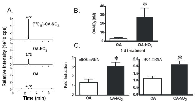
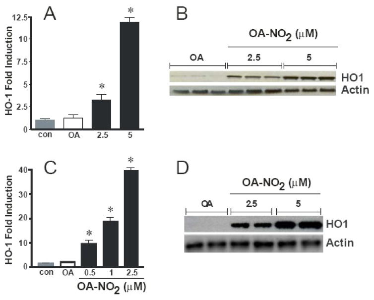
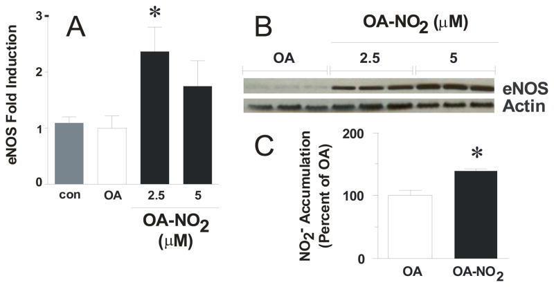
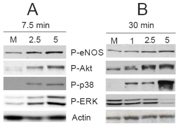
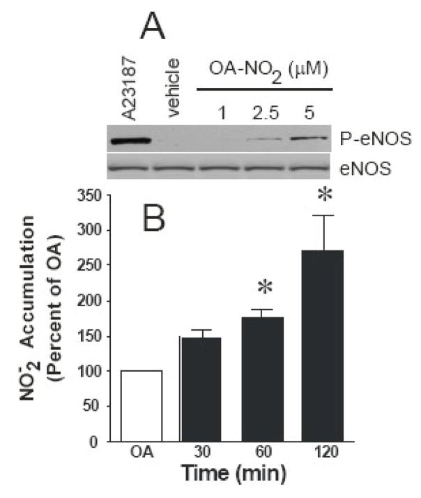
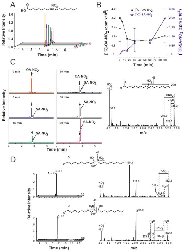
Reactive oxygen species mediate a decrease in nitric oxide (NO) bioavailability and endothelial dysfunction, with secondary oxidized and nitrated by-products of these reactions contributing to the pathogenesis of numerous vascular diseases. While oxidized lipids and lipoproteins exacerbate inflammatory reactions in the vasculature, in stark contrast the nitration of polyunsaturated fatty acids and complex lipids yields electrophilic products that exhibit pluripotent anti-inflammatory signaling capabilities acting via both cGMP-dependent and -independent mechanisms. Herein we report that nitro-oleic acid (OA-NO(2)) treatment increases expression of endothelial nitric oxide synthase (eNOS) and heme oxygenase 1 (HO-1) in the vasculature, thus transducing vascular protective effects associated with enhanced NO production. Administration of OA-NO(2) via osmotic pump results in a significant increase in eNOS and HO-1 mRNA in mouse aortas. Moreover, HPLC-MS/MS analysis showed that NO(2)-FAs are rapidly metabolized in cultured endothelial cells (ECs) and treatment with NO(2)-FAs stimulated the phosphorylation of eNOS at Ser(1179). These posttranslational modifications of eNOS, in concert with elevated eNOS gene expression, contributed to an increase in endothelial NO production. In aggregate, OA-NO(2)-induced eNOS and HO-1 expression by vascular cells can induce beneficial effects on endothelial function and provide a new strategy for treating various vascular inflammatory and hypertensive disorders.


Figures

Figure 1
Figure 1
OA-NO2 levels increased in mice treated subcutaneously with OA or OA-NO2 via osmotic mini pump for three days accompanies enhanced eNOS and HO-1 gene expression. Blood was collected from the treated mice. The serum was analyzed by HPLC ESI MS/MS in the negative ion mode using [13C]OA-NO2 as an internal standard by acquiring MRM transitions consistent with the loss of the nitro functional group (46 amu corresponds to the mass of NO2): m/z 326/46 and m/z 344/46 for OA-NO2 and [13C]OA-NO2 respectively. Representative chromatographs of lipids extracted from serum [13C18] OA-NO2 (top panel), OA-NO2 (middle panel), and OA (bottom panel) are shown (A). Free OA-NO2 levels (nM) were determined using ANALYST 1.4 quantitation software (B). Real time PCR analysis was performed for eNOS and HO-1 from aortas of mice with the osmotic mini pump for 3 d. Administration of OA-NO2 (3 mg/kg/d for 3 d) increased eNOS (left) and HO-1(right) mRNA levels compared to OA-treated mice. eNOS and HO-1 mRNA levels were normalized to Actin (C). Data are expressed from 6–8 mice per group and expressed as mean ± SEM.
Figure 2
Figure 2
OA-NO2 induces HO-1 mRNA and protein in endothelial and vascular smooth muscle cells. Endothelial cells were incubated with 2.5 and 5 μM OA-NO2 or the native fatty acid OA for 16 hrs and real time PCR analysis was performed (A). Endothelial cells were treated with the same concentrations of OA-NO2 or OA for 24 hr and Western blotting was performed (B). VSMCs were treated with the indicated concentrations of OA-NO2 or OA for 2.5 hr and 24 hr and real time PCR analysis (C) and Western blotting (D) was performed, respectively. The Western blot is a representative of 5–7 separate experiments. The real time PCR analysis results are derived from at least five independent experiments and data are expressed as mean ± SEM.
Figure 3
Figure 3
Endothelial cells increase eNOS following OA-NO2 treatment. Cells were incubated for 16 hr with indicated concentrations of OA-NO2 or OA and real time PCR analysis was performed (A) and 24 hr for Western blotting (B). OA-NO2 treatment for 16 hr stimulates NO release from ECs. NO generation was determined in OA- and 2.5 μM OA-NO2-treated confluent ECs for 16 hrs using Sievers nitric oxide analyzer (NOA). The levels of nitrite accumulation have been normalized to protein content of the ECs and are reported as a percent of the OA control. Basal levels of NO2 = 326.5 ± 8.7 pmoles/mg protein. Results are derived from at least five independent experiments and data are expressed as mean ± SEM.
Figure 4
Figure 4
OA-NO2 treatment stimulates phosphorylation of eNOS, Akt, ERK, and p38. Confluent ECs were stimulated with 2.5 and 5 μM OA-NO2 for 7.5 (A) and 30 min (B) and Western blotted for P-eNOS, P-Akt, P-p38, P-ERK, and actin. The results are a representative of 5–7 separate experiments.
Figure 5
Figure 5
OA-NO2 treatment stimulates NO release from ECs. NO generation was determined in OA- and 2.5 μM OA-NO2-treated confluent ECs for the indicated times using Sievers nitric oxide analyzer (NOA). The levels of nitrite have been normalized to protein concentration (basal NO2 = 4.8 ± 0.2 pmoles/min/mg protein) and are reported as a percent of control at each individual time point (30, 60, and 120 min). Results are derived from at least five independent experiments and data are expressed as mean ± SEM.
Figure 6
Figure 6
OA-NO2 is rapidly metabolized by ECs. BAEC were grown to confluence and treated with a mixture of 9- and 10-nitro regio-isomers of [13C18]-OA-NO2 (5 μM) in HBSS over a period of 90 minutes. Equimolar distribution of the 9- and 10-nitro regio-isomers of OA-NO2 is depicted (A, inset) as previously determined ([7], online supplement). The HBSS solution was collected and ECs were scrape harvested at the indicated time points. HBSS, corresponding cell extracts and control HBSS treatments (with [13C18]-OA-NO2 at 37°C at each indicated time point) were extracted using ACN precipitation as described in Materials and Methods and supernatants were analyzed by LC-MS-MS. [13C18]-OA-NO2 was detected following a m/z 344/46 for [13C18]-OA-NO2 and a MRM transition corresponding to the detection of the nitro group (NO2) with peak at 3.6 min. A continuous decay in peak area was observed for [13C18]-OA-NO2 over the 90 min time course (A). A composite of the chromatograms shows the loss of the [13C18]-OA-NO2 (filled square) and the concomitant formation of [13C18]-SA-NO2 (open triangle) over the period of 90 min (B). [13C18]-OA-NO2 is rapidly saturated to a nitro-stearic acid ([13C18]-SA-NO2). [13C18]-SA-NO2 was detected following a m/z 346/46 MRM transition (C, left panel). Arrow indicates a peak corresponding to the contribution of heavy isotopes from [13C18]-OA-NO2. Fragmentation patterns of product ion analysis (peak at 3.73 min) confirmed the formation of [13C18]-SA-NO2. The major expected fragments from [13C18]-SA-NO2 were detected, corresponding to the formation of 46 (NO2): m/z 299 (neutral loss of HNO2,-47 amu) and 328 (the loss H2O, −18 amu) (C, right panel). Scheme showing the main fragmentation products (C, inset right panel). The two isomers of [13C18]-OA-(OH)-NO2 were detected following a formation of specific breakdown product ions. The MRM transition m/z 362/180 corresponds to the fragmentation of 9-(OH)-10NO2 (peak at 5.7 and 5.8 min) (D, upper panels), and transition m/z 362/211 corresponds to the fragmentation of 10-(OH)-9NO2 (peak at 5.8 min) (D, lower panels). Fragmentation patterns of [13C18]-OA-(OH)-NO2 isomers (D, right panels) show characteristic fragmentation for vicinal nitro-hydroxy fatty acids (180 and 193 ions) and formation of NO2 (46 ion).

References

    1. Schopfer FJ, Baker PR, Giles G, Chumley P, Batthyany C, Crawford J, Patel RP, Hogg N, Branchaud BP, Lancaster JR, Jr, Freeman BA. Fatty acid transduction of nitric oxide signaling. Nitrolinoleic acid is a hydrophobically stabilized nitric oxide donor. J Biol Chem. 2005;280:19289–19297. - PubMed
    1. Baker LM, Baker PR, Golin-Bisello F, Schopfer FJ, Fink M, Woodcock SR, Branchaud BP, Radi R, Freeman BA. Nitro-fatty acid reaction with glutathione and cysteine. Kinetic analysis of thiol alkylation by a Michael addition reaction. J Biol Chem. 2007;282:31085–31093. - PMC - PubMed
    1. Batthyany C, Schopfer FJ, Baker PR, Duran R, Baker LM, Huang Y, Cervenansky C, Branchaud BP, Freeman BA. Reversible post-translational modification of proteins by nitrated fatty acids in vivo. J Biol Chem. 2006;281:20450–20463. - PMC - PubMed
    1. Cui T, Schopfer FJ, Zhang J, Chen K, Ichikawa T, Baker PR, Batthyany C, Chacko BK, Feng X, Patel RP, Agarwal A, Freeman BA, Chen YE. Nitrated fatty acids: Endogenous anti-inflammatory signaling mediators. J Biol Chem. 2006;281:35686–35698. - PMC - PubMed
    1. Baker PR, Lin Y, Schopfer FJ, Woodcock SR, Groeger AL, Batthyany C, Sweeney S, Long MH, Iles KE, Baker LM, Branchaud BP, Chen YE, Freeman BA. Fatty acid transduction of nitric oxide signaling: multiple nitrated unsaturated fatty acid derivatives exist in human blood and urine and serve as endogenous peroxisome proliferator-activated receptor ligands. J Biol Chem. 2005;280:42464–42475. - PMC - PubMed

Publication types

MeSH terms