. 2009 Nov;11(11):891-900.
doi: 10.1016/j.jfms.2009.09.010.

Brachycephalic feline noses: CT and anatomical study of the relationship between head conformation and the nasolacrimal drainage system

Affiliations

Brachycephalic feline noses: CT and anatomical study of the relationship between head conformation and the nasolacrimal drainage system

Claudia Schlueter et al. J Feline Med Surg. 2009 Nov.

Abstract

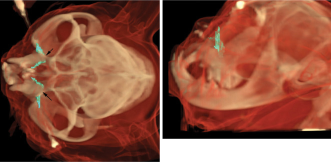
Aims: A study was designed to evaluate the influence of head conformation on the course of the nasolacrimal drainage system (NDS) in 31 brachycephalic and 15 mesocephalic cats using computed tomography (CT), CT-dacryocystography and anatomical methods.

Findings: The higher the degree of brachycephalia, the more the facial bones and upper canine teeth are displaced dorsally (ie, the more pronounced the dorsorotation). Dorsorotation leads to abnormal dislocation of the ventral nasal concha and to almost horizontally rotated upper canine teeth, and thus a steeply oriented NDS. In severe brachycephalia the NDS is forced to pass below the canine tooth (adopt a V-shaped course) and the drainage function seems to be inefficient.

Practical relevance: The rotation of the upper canine teeth appears to provide a basis for classification of brachycephalia in cats. The authors recommend that breeders avoid breeding from individuals affected by this condition and to give preference to cats with longer facial bones.


Figures

FIG 1
FIG 1
Radiographs of a mesocephalic cat (a) and a severely brachycephalic cat (Exotic shorthair) (b). Note the length and the tip of the nose and nasal bone, the dorsally rotated hard palate, nasal airways and jaw, the compressed and dislocated conchae, and the elongated and thickened soft palate in the brachycephalic cat
FIG 2
FIG 2
To account for the range in phenotypic appearance, a classification system with four degrees of brachycephalia - from mild (I) to severe (IV) - is recommended. The main features on which this classification is based are the dorsal displacement of the maxillary canine teeth and the dorsorotation of the jaw. Note that drainage discoloration of the skin starts in moderately affected cats; and that the tip of the nose is located at a higher level than the lower eyelid in profound and severe degrees of brachycephalia
FIG 3
FIG 3
In the background a skull of a domestic cat and a brachycephalic cat (category II, moderate) are illustrated. Superimposed on these is a skull with a severe (category IV) degree of brachycephalia. Note the highly shortened facial skull, and the cumulative effects of this reduction within the area of the nose. There is no nasal bone and reduced maxillary bone, and it would seem impossible for all teeth to be accommodated. The viscerocranium and the mandible are dorsally rotated also, and the canine tooth is almost horizontal in position. The whole lacrimal bone is reduced to a prominent process (processus frontalis of the lacrimal bone). Drawing courtesy of Gisela Jahrmärker
FIG 4
FIG 4
The ventral nasal bone (VNB) of a mesocephalic cat (left) and brachycephalic cat (category II, moderate) (right) is displayed from a dorsomedial view; the conchae ethmoidales and nasal septum have been removed. The maxillary canine tooth markedly influences the course of the nasolacrimal drainage (marked in red). Compared with mesocephalic cats, the VNB in brachycephalic cats is dorsally rotated (mobile point). The drainage streams adjacent to the basal lamina of the ventral nasal bone (1) and is therefore also characterised by an increased angle and a steeper course
FIG 5
FIG 5
Comparison of transverse contrast CT images (A and C) and transverse non-contrast CT images (B and D) of moderate (category II) brachycephalia. The cast within the skull of this cat displays the course of the NDS and its topographic location compared with the canine tooth. The distance between the root of the tooth (indicated by the dashed line) and the nasolacrimal sac is 2 mm. Lateral to the processus frontalis of the lacrimal bone (2a) is the upper lacrimal canaliculus (1a). 1b = lower lacrimal canaliculus, 1c = lacrimal sac, 2b = fossa sacci lacrimalis, 1d = nasolacrimal duct
FIG 6
FIG 6
Comparison of a transverse contrast CT image (left) and a transverse non-contrast CT image (right) of severe (category IV) brachycephalia. To drain from the lacrimal sac (1c) into the nose, the nasolacrimal duct (1d) is forced to pass below the root of the canine tooth (c). The root of the canine tooth (indicated by a dashed line) is situated directly adjoining the nasolacrimal sac (1c). Note the prominent frontal process of the lacrimal bone in the left-hand image (2a). The cast within the skull of this cat is hidden by the canine tooth root
FIG 7
FIG 7
Three-dimensional models of the NDS in moderate brachycephalia; dorsoventral and laterolateral views. An acute angle and caudal-to-rostral course is visible. Arrows mark the position of the canine teeth
FIG 8
FIG 8
Three-dimensional models of the NDS in severe brachycephalia demonstrate the V-shaped course and reveal what is occult in the cast. Arrows mark the location of the canine teeth
FIG 9
FIG 9
Three-dimensional model of a mesocephalic cat. Note the course of the nasolacrimal duct, which runs parallel to the hard palate and shows a right-angled alignment
None

Comment in

References

    1. Fournier PF. The Lorenz theory of beauty. J Cosmet Dermatol 2002; 1: 131–36. - PubMed
    1. Künzel W, Breit S, Oppel M. Morphometric investigations of breed-specific features in feline skulls and considerations on their functional implications. Anat Histol Embryol 2003; 32: 218–23. - PubMed
    1. Lauruschkus G. Über riechfeldgröße und riechfeldkoeffizient bei einigen hunderassen und der katze. Archiv für Tierheilkunde 1942; 77: 473–97.
    1. Hobson HP. Brachycephalic syndrome. Semin Vet Med Surg (Small Anim) 1995; 10:109–14. - PubMed
    1. Barnett KC, Crispin SM. Feline ophthalmology. London: WB Saunders; 1998.

MeSH terms