Review
. 2009 Dec;13(5-6):678-86.
doi: 10.1016/j.cbpa.2009.09.026. Epub 2009 Oct 24.

Light activation as a method of regulating and studying gene expression

Affiliations
Review

Light activation as a method of regulating and studying gene expression

Alexander Deiters. Curr Opin Chem Biol. 2009 Dec.

Abstract

Recently, several advances have been made in the activation and deactivation of gene expression using light. These developments are based on the application of small molecule inducers of gene expression, antisense- or RNA interference-mediated gene silencing, and the photochemical control of proteins regulating gene function. The majority of the examples employ a classical 'caging technology', through the chemical installation of a light-removable protecting group on the biological molecule (small molecule, oligonucleotide, or protein) of interest and rendering it inactive. UV light irradiation then removes the caging group and activates the molecule, enabling control over gene activity with high spatial and temporal resolution.


Figures

Figure 1
Figure 1
General decaging reaction, and a classical ortho-nitrobenzyl caging group. Chemical and photochemical properties of the caging group can be tuned with suitable substituents, including: R1 = H, R2 = H; R1 = OCH3, R2 = H; R1 = OCH3, R2 = CH3. Adapted with permission the Royal Society of Chemistry from: Org. Biomol. Chem. 2007, 5:999–1005.
Figure 2
Figure 2
Light-activated small molecule inducers of gene expression, caged doxycycline (1) and caged toyocamycin (2). The caging group (blue) is removed through UV irradiation.
Figure 3
Figure 3
Photoactivated gene expression in a mouse embryo. (a) Fluorescence image of a control embryonic day 10.5 (E10.5) embryo (CIG-tGFP) incubated for 24 h with 20 μM doxycycline, displaying widespread GFP fluorescence particularly in neurons of the spinal cord dorsal root ganglia, the developing heart and in trigeminal ganglia of the head. (b) Fluorescence image of an E10.5 embryo incubated with 2.6 μM of 1 without irradiation. (c) E10.5 embryo after a single 15 sec pulse of UV light, and (d) after a second pulse of irradiation 3 h later. (e) Spatially restricted fluorescence in three dorsal root ganglia of an E10.5 embryo after localized irradiation at the tip of the tail with a spot size of about three ganglia (circle). Adapted by permission from Macmillan Publishers Ltd: Nat. Methods 2009, 6:527–531, copyright 2009.
Figure 4
Figure 4
A functional hammerhead ribozyme located in the 5′ UTR of a gene leads to gene silencing through 5′ cap removal. The irradiation of 2 leads to toyocamycin formation which inhibits ribozyme function and induces gene expression (e.g. luciferase or GFP). This enables spatial control of GFP activation in a monolayer of HEK293T cells. The area within the white circle was irradiated with UV light (360 nm, 30 sec), and the cells were imaged after 24 h to allow for GFP expression and maturation. Adapted with permission of the Royal Society of Chemistry from: Chem. Commun. 2009, 568–570.
Figure 5
Figure 5
Two different approaches to the photochemical regulation of gene downregulation via antisense agents/RNA interference.
Figure 6
Figure 6
Caged thymidine nucleotide 3. The caging group (blue) is removed through UV irradiation. a) Spatial regulation of Renilla luciferase expression using caged SDNA antisense agents. The cellular monolayer was only irradiated inside the white circle (365 nm, 5 min, 23 W), leading to localized silencing of luciferase in cells transfected with the caged SDNA. b) Cells which have been locally irradiated but not been transfected with the caged SDNA do not show luciferase silencing. Adapted with permission from: ChemBioChem 2008, 9: 2937–2940. Copyright Wiley-VCH Verlag GmbH & Co. KGaA.
Figure 7
Figure 7
Base- and phosphate-caged nucleotides 4 and 56, respectively. The caging group (blue) is removed through UV irradiation.
Figure 8
Figure 8
Zebrafish embryos at 28 hours past fertilization (hpf); the box indicates the region in which image analysis was performed. Caged and control siFNA were co-injected with a GFP expression plasmid. Exposure to UV light (365 nm) was performed at 7 hpf, and photographs were taken at 28 hpf. Caged siFNA injections resemble control GFP mosaic expressions, but light exposure activated the siFNAs and resulted in the silencing of GFP expression. Adapted with permission of the Royal Society of Chemistry from: Mol. Biosyst. 2008, 4:431–440.
Figure 9
Figure 9
Zebrafish embryos at 24 hpf. a) Ntl null mutant displaying a characteristic phenotype with a greatly reduced tail and misshaped somites. b) An embryo injected with an anti-ntl MO shows the same phenotype, while c) an embryo injected with a caged anti-ntl MO shows a wild-type phenotype. d) Irradiation of embryos injected with the caged MO at the sphere stage results in a clean ntl mutant phenotype, thus validating light-activation of gene silencing. Adapted by permission from Macmillan Publishers Ltd: Nat. Chem. Biol. 2007, 3:650–651, copyright 2007.
Figure 10
Figure 10
Cre recombinase was rendered inactive through site-specific incorporation of an ortho-nitrobenzyl protected tyrosine residue at position Y324. Irradiation with UV light (365 nm) restores activity and triggers DNA recombination. a) HEK293T cells uniformly expressing DsRed. b) Transfection of wild-type Cre recombinase leads to the excision of the DsRed gene and the activation of GFP expression. c) Caged Cre recombinase is completely inactive, until d) irradiated with UV light (365 nm, 5 min), thus catalyzing DNA recombination. Adapted with permission from: ACS Chem. Biol. 2009, 4:441–445. Copyright 2009 American Chemical Society
Figure 11
Figure 11
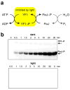
Kinase activity of fusion kinase YF1. a) The YF1 autophosphorylation and then transfer of the phosphoryl group to its substrate, the response regulator FixJ, is inhibited by light irradiation. b) This photochemical switching of activity is visualized on two gels which display a time course of FixJ phosphorylation with [γ-32P] ATP, catalyzed by YF1. Adapted from: J. Mol. Biol. 2008, 385:1433–1444, with permission from Elsevier.
Figure 12
Figure 12
a) Reversible light-regulation of the Rac1-LOV fusion protein. b) HeLa cells expressing Rac1-LOV showed lamellipodial protrusions and membrane ruffles within minutes after light irradiation. Adapted by permission from Macmillan Publishers Ltd: Nature 2009 461:104–108, copyright 2009
Figure 13
Figure 13
a) Relationship between induced cellular temperature and power of the IR laser 5 s after the initiation of irradiation. b) Infrared-laser irradiation (11 mW, 1 s) induced cytoplasmic GFP expression, under control of a heat-shock promoter, in the targeted seam cell (arrow) of an L4 larva 3 h after irradiation. Adapted by permission from Macmillan Publishers Ltd: Nat. Methods 2009, 6:79–81, copyright 2009.

References

    1. Casey JP, Blidner RA, Monroe WT. Caged siRNAs for Spatiotemporal Control of Gene Silencing. Mol Pharm. 2009;6:669–685. - PubMed
    1. Young DD, Deiters A. Photochemical control of biological processes. Org Biomol Chem. 2007;5:999–1005. - PubMed
    1. Tang X, Dmochowski IJ. Regulating gene expression with light-activated oligonucleotides. Mol Biosyst. 2007;3:100–110. - PubMed
    1. Mayer G, Heckel A. Biologically active molecules with a “light switch”. Angew Chem Int Ed. 2006;45:4900–4921. - PubMed
    1. Curley K, Lawrence DS. Light-activated proteins. Curr Opin Chem Biol. 1999;3:84–88. - PubMed

Publication types

LinkOut - more resources