. 2010 Mar;24(3):740-9.
doi: 10.1096/fj.09-144147. Epub 2009 Oct 26.

Increased adenosine contributes to penile fibrosis, a dangerous feature of priapism, via A2B adenosine receptor signaling

Affiliations

Increased adenosine contributes to penile fibrosis, a dangerous feature of priapism, via A2B adenosine receptor signaling

Jiaming Wen et al. FASEB J. 2010 Mar.

Abstract

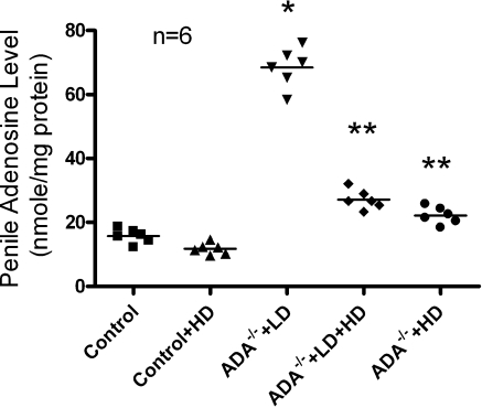
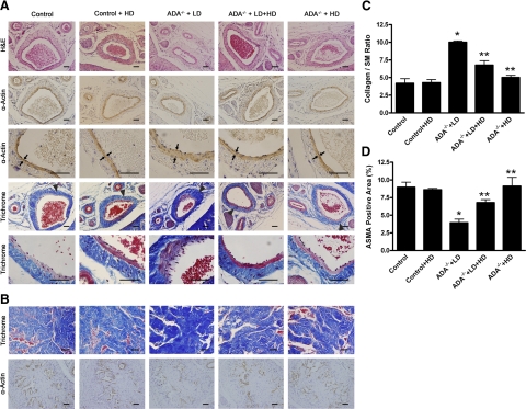
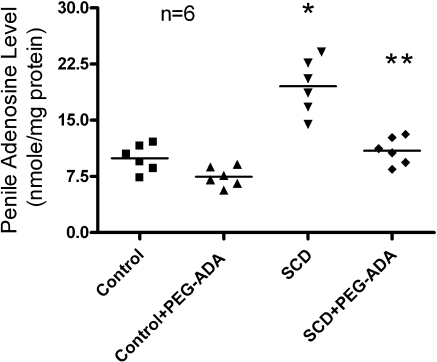
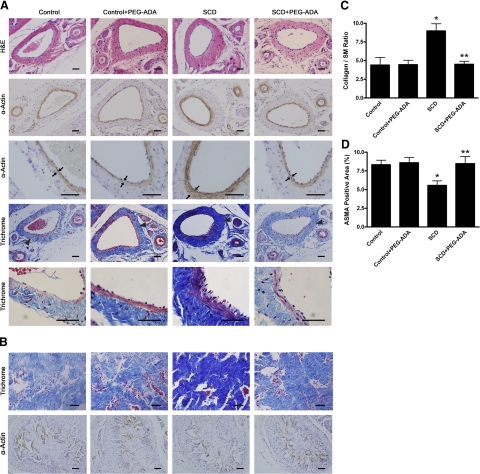
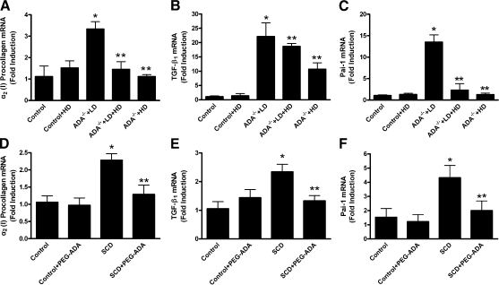
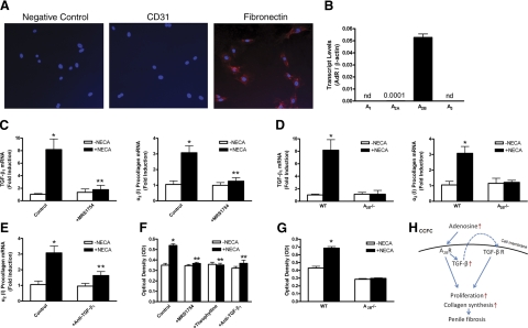
Priapism is a condition of persistent penile erection in the absence of sexual excitation. Of men with sickle cell disease (SCD), 40% display priapism. The disorder is a dangerous and urgent condition, given its association with penile fibrosis and eventual erectile dysfunction. Current strategies to prevent its progression are poor because of a lack of fundamental understanding of the molecular mechanisms for penile fibrosis in priapism. Here we demonstrate that increased adenosine is a novel causative factor contributing to penile fibrosis in two independent animal models of priapism, adenosine deaminase (ADA)-deficient mice and SCD transgenic mice. An important finding is that chronic reduction of adenosine by ADA enzyme therapy successfully attenuated penile fibrosis in both mouse models, indicating an essential role of increased adenosine in penile fibrosis and a novel therapeutic possibility for this serious complication. Subsequently, we identified that both mice models share a similar fibrotic gene expression profile in penile tissue (including procollagen I, TGF-beta(1), and plasminogen activator inhibitor-1 mRNA), suggesting that they share similar signaling pathways for progression to penile fibrosis. Thus, in an effort to decipher specific cell types and underlying mechanism responsible for adenosine-mediated penile fibrosis, we purified corpus cavernosal fibroblast cells (CCFCs), the major cell type involved in this process, from wild-type mice. Quantitative RT-PCR showed that the major receptor expressed in these cells is the adenosine receptor A(2B)R. Based on this fact, we further purified CCFCs from A(2B)R-deficient mice and demonstrated that A(2B)R is essential for excess adenosine-mediated penile fibrosis. Finally, we revealed that TGF-beta functions downstream of the A(2B)R to increase CCFC collagen secretion and proliferation. Overall, our studies identify an essential role of increased adenosine in the pathogenesis of penile fibrosis via A(2B)R signaling and offer a potential target for prevention and treatment of penile fibrosis, a dangerous complication seen in priapism.-Wen, J., Jiang, X., Dai, Y., Zhang, Y., Tang, Y., Sun, H., Mi, T., Phatarpekar, P. V., Kellems, R. E., Blackburn, M. R., Xia, Y. Increased adenosine contributes to penile fibrosis, a dangerous feature of priapism, via A(2B) adenosine receptor signaling.


Figures

Figure 1.
Figure 1.
Modulation of penile adenosine levels in ADA-deficient mice after different dosages of PEG-ADA treatment. Data are expressed as means ± se (n=6). *P < 0.05 vs. control; **P < 0.05 vs. ADA−/− + LD.
Figure 2.
Figure 2.
Elevated adenosine level in penile tissue contributes to penile fibrosis in ADA-deficient mice. A) For LD PEG-ADA-treated ADA-deficient mice, H&E staining and anti-α-SMA immunostaining demonstrated extensive endothelial damage, including marked intimal thickening with smooth muscle hypertrophy and endothelial swelling in the deep dorsal vein. Masson’s trichrome staining showed significant fibrosis with extension into the intima that was also seen in the deep dorsal vein. Vascular damage and penile fibrosis were prevented in the HD-treated mice since their birth (HD group) and attenuated with HD PEG-ADA treatment after establishment of penile fibrosis in LD-treated mice (LD+HD group), respectively. Arrows indicate hypertrophy of smooth muscle of the deep dorsal vein in LD-treated mice. Triangles indicate vascular damage featured with intimal thickening and fibrosis in LD-treated mice. B) Masson’s trichrome staining and anti-α-SMA immunostaining showed substantial fibrosis in the corpus cavernosum accompanied by loss of vascular smooth muscle cells in ADA-deficient mice receiving the LD treatment regimen. These features were either prevented or attenuated in mice receiving the HD treatment since birth or the LD + HD PEG-ADA treatment regimen, respectively. C, D) Quantitative image analysis showed that LD PEG-ADA-treated mice exhibited an increased collagen to smooth muscle (SM) ratio (C) and decreased positive α-actin smooth muscle area (ASMA) in corpus cavernosum (D). Data are expressed as means ± se (n=5). *P < 0.05 vs. control; **P < 0.05 vs. ADA−/− + LD. Scale bars = 50 μm.
Figure 3.
Figure 3.
Elevated adenosine levels in penile tissues of SCD-Tg mice were reduced by PEG-ADA enzyme therapy. Data are expressed as means ± se (n=6). *P < 0.05 vs. control; **P < 0.05 vs. SCD.
Figure 4.
Figure 4.
SCD-Tg mice display penile fibrosis due to elevations of adenosine levels in penile tissues. A) Very similar to ADA-deficient mice, SCD-Tg mice without PEG-ADA treatment displayed vascular damage and fibrosis in the deep dorsal vein. Lowering penile adenosine levels after established penile fibrosis by PEG-ADA enzyme therapy resulted in a significant resolution of vascular damage and fibrosis. Arrows indicate hypertrophy of smooth muscle in the deep dorsal vein of SCD mice. Triangles indicate penile vascular damage featured with intimal thickening and fibrosis in SCD mice. B) SCD-Tg mice exhibited penile fibrosis and smooth muscle damage in the corpus cavernosum. Lowering penile adenosine levels after established priapism and fibrosis by PEG-ADA enzyme therapy attenuates these abnormalities. C, D) Quantitative image analyses showed that SCD-Tg mice exhibited the increased collagen to smooth muscle (SM) ratio (C) and decreased positive α-actin smooth muscle area (ASMA) in the corpus cavernosum (D). Data are expressed as means ± se (n=6). *P < 0.05 vs. control; **P < 0.05 vs. SCD. Scale bars = 50 μm.
Figure 5.
Figure 5.
Elevated adenosine contributes to increased expression of fibrotic marker genes in the penile tissues of LD-treated ADA-deficient mice and SCD-Tg mice. A–C) Expression of the fibrotic marker genes in penile tissues of ADA-deficient mice receiving the LD regimen significantly increased. HD and LD+HD PEG-ADA treatment regimens for ADA-deficient mice prevented and attenuated the increased expression of fibrotic mediators. Collagen I mRNA (A), TGF-β1 mRNA (B), and PAI-1 mRNA (C) in control and PEG-ADA-treated mice. Data are expressed as means ± se (n=4–5). *P < 0.05 vs. control; **P < 0.05 vs. ADA−/− + LD. D–F) SCD-Tg mice exhibited increased expression of fibrotic mediators in penile tissue. PEG-ADA inhibited the increased expression in SCD-Tg mice. Collagen I mRNA (D) TGF-β1 mRNA (E), and PAI-1 mRNA (F). Data are expressed as means ± se (n=5–6). *P < 0.05 vs. control; **P < 0.05 vs. SCD.
Figure 6.
Figure 6.
TGF-β is a common intracellular signaling molecule downstream of the A2B adenosine receptor responsible for excess adenosine-mediated penile fibrosis by increased procollagen production and proliferation of fibroblasts. A) Primary isolated CCFCs were stained by fibronectin, a fibroblast-specific marker, not by CD 31, an endothelial cell-specific marker. B) A2BR is major receptor expressed in CCFCs. Quantitative RT-PCR was used to determine the gene expression of adenosine receptors in CCFCs. Data are expressed as means ± se (n=4). C) NECA increased the expression of TGF-β1 mRNA and procollagen I mRNA levels in CCFCs, which was completely abolished by the A2BR specific antagonist, MRS1754 (n=4–6). *P < 0.05 vs. no treatment; **P < 0.05 vs. NECA treatment. D) Genetic deletion of A2BR abolished the NECA-induced procollagen I and TGF-β1 mRNA production (n=5). *P < 0.05 vs. untreated wild-type CCFCs. E) NECA-induced procollagen I mRNA expression was abolished by TGF-β1-neutralizing antibody (n=4–6). *P < 0.05 vs. no treatment; **P < 0.05 vs. NECA treatment. F) Adenosine-induced TGF-β production leads to increased fibroblast proliferation via A2BR activation. NECA, a nonmetabolized adenosine receptor agonist (20 μM), significantly increased CCFC proliferation. MRS1754, a A2BR specific antagonist (20 μM), theophylline, a general adenosine receptor antagonist (20 μM), and TGF-β1-neutralizing antibody (0.5 μg/ml) markedly inhibited NECA-induced proliferation. Data are expressed as means ± se (n=8). *P < 0.05 vs. no treatment; **P < 0.05 vs. NECA treatment. G) Genetic deletion of A2BR completely abolished NECA-induced fibroblast proliferation (n=8). *P < 0.05 vs. untreated wild-type CCFCs. H) Signaling pathway for excess adenosine-mediated penile fibrosis. Excess adenosine increases the expression of TGF-β1 via A2B adenosine receptor in fibroblasts. Increased TGF-β1 functioning as an autocrine fashion leads to penile fibrosis by increased collagen synthesis and proliferation in fibroblasts in the corpus cavernosum.

References

    1. Bruno D, Wigfall D R, Zimmerman S A, Rosoff P M, Wiener J S. Genitourinary complications of sickle cell disease. J Urol. 2001;166:803–811. - PubMed
    1. Diggs L W, Ching R E. Pathology of sickle cell anemia. South Med J. 1934;27:839–845.
    1. Van der Horst C, Stuebinger H, Seif C, Melchior D, Martinez-Portillo F J, Juenemann K P. Priapism—etiology, pathophysiology and management. Int Braz J Urol. 2003;29:391–400. - PubMed
    1. Burnett A L. Erectile dysfunction. J Urol. 2006;175:S25–31. - PubMed
    1. Dai Y, Zhang Y, Phatarpekar P, Mi T, Zhang H, Blackburn M R, Xia Y. Adenosine signaling, priapism and novel therapies. J Sex Med. 2009;6:292–301. - PubMed

Publication types

MeSH terms

LinkOut - more resources