. 2010 Jan;30(1):284-94.
doi: 10.1128/MCB.00651-09.

Tom20 mediates localization of mRNAs to mitochondria in a translation-dependent manner

Affiliations

Tom20 mediates localization of mRNAs to mitochondria in a translation-dependent manner

Erez Eliyahu et al. Mol Cell Biol. 2010 Jan.

Abstract

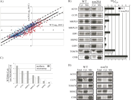
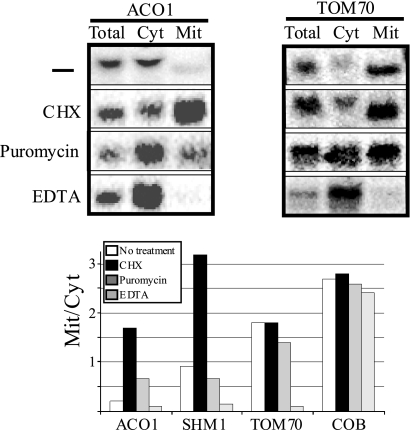
mRNAs encoding mitochondrial proteins are enriched in the vicinity of mitochondria, presumably to facilitate protein transport. A possible mechanism for enrichment may involve interaction of the translocase of the mitochondrial outer membrane (TOM) complex with the precursor protein while it is translated, thereby leading to association of polysomal mRNAs with mitochondria. To test this hypothesis, we isolated mitochondrial fractions from yeast cells lacking the major import receptor, Tom20, and compared their mRNA repertoire to that of wild-type cells by DNA microarrays. Most mRNAs encoding mitochondrial proteins were less associated with mitochondria, yet the extent of decrease varied among genes. Analysis of several mRNAs revealed that optimal association of Tom20 target mRNAs requires both translating ribosomes and features within the encoded mitochondrial targeting signal. Recently, Puf3p was implicated in the association of mRNAs with mitochondria through interaction with untranslated regions. We therefore constructed a tom20 Delta puf3 Delta double-knockout strain, which demonstrated growth defects under conditions where fully functional mitochondria are required. Mislocalization effects for few tested mRNAs appeared stronger in the double knockout than in the tom20 Delta strain. Taken together, our data reveal a large-scale mRNA association mode that involves interaction of Tom20p with the translated mitochondrial targeting sequence and may be assisted by Puf3p.


Figures

FIG. 1.
FIG. 1.
Analysis of mitochondrial and cytosolic fractions. Yeast cells were treated with zymolase and lysed with a Dounce homogenizer. Cellular compartments were fractionated to crude mitochondrial (Mit) or cytosolic (Cyt) fractions by sequential centrifugation. (A) RNA from the cytosolic or mitochondrial fractions was subjected to Northern analyses with probes recognizing mitochondrially encoded RNAs (15S rRNA and COB) or nuclear-encoded mRNAs (SEC61 and SHM2). The Total lane includes RNA that was collected before fractionation, and a one-third equivalent of the fractionated RNA was loaded. The signal in the Total lane was therefore usually one-third of the sum of mitochondrial and cytosolic signals. (B) Subcellular fractions of wild-type (WT) and tom20Δ cells were analyzed by Western blotting. Antibodies recognizing markers for the mitochondria (Aco1 and Tom20), nuclear-membrane (Npc), cytosol (Hxk1), ER (Sec61 and Bip1), and plasma membrane (Gas1) were used. Equal amounts of unfractionated protein sample (Total) were included as controls to demonstrate that there are no significant losses during fractionation. Note that the two bands that appear in the cytosolic fraction of Tom20p are nonspecific signals because they appear also in the tom20Δ strain upon longer exposure (not shown).
FIG. 2.
FIG. 2.
TOM20 deletion specifically affects mitochondrial association of hundreds of mRNAs. (A) Scatter plot where each spot indicates the relative amount of each mRNA in the mitochondrial fraction in the parental strain (wild type [WT]) and the tom20Δ cells. The solid line indicates the best-fit linear trend line, and the two dashed lines represent 2 SDs above and below the trend line. Red spots indicate genes that are designated mitochondrial in the SGD, and blue spots represent all other genes. (B) Northern analyses of the change in distribution of the indicated mRNAs in the wild-type and tom20Δ cells. Bars represent the ratio of the M/C signal in the WT and tom20Δ cells. (C) Comparison of the change in mitochondrial association obtained by the microarray analysis and the Northern analysis. (D) Northern analyses of the distribution of the indicated mRNAs in the wild-type and tom7Δ cells. Lanes are as described in the legend to Fig. 1A.
FIG. 3.
FIG. 3.
Association of Tom20 target mRNAs is translation dependent. Cells were grown in a medium containing galactose and subjected to fractionation in the absence or presence of polyribosome stabilizer (CHX) or destabilizers (puromycin) or with a divalent ion chelator (EDTA). RNA samples were subjected to Northern analysis using probes that recognize Tom20 target mRNAs (ACO1 and SHM1), non-Tom20 targets (TOM70), and mitochondrially transcribed mRNA (COB). Lane loading is as described in the legend of Fig. 1. Northern blots for two representative mRNAs and a bar chart of quantitation of all mRNAs are shown.
FIG. 4.
FIG. 4.
mRNA association with mitochondria is MTS-dependent. (A) Wild-type (WT) or tom20Δ cells were transformed with a plasmid expressing either a GFP reporter alone (GFP) or GFP fused to the Su9 MTS (MTS-GFP), to the BCS1 3′ UTR (GFP-3′ UTR), or to the MTS and the 3′ UTR (MTS-GFP-3′ UTR). Cells were fractionated, and RNA samples were subjected to Northern analysis with probe recognizing GFP. The bar chart presents the quantitation of the data from the wild-type or tom20Δ cells. Error bars indicate the standard errors of the means where at least three experiments were performed. (B) Plasmids expressing either the normal ACO1 coding region (WT) or ACO1 variants (H, J, and L) carrying point mutations in the MTS (indicated in bold) were introduced into aco1Δ cells. Cells were fractionated, and the M/C signal ratio is shown (average of three experiments). The number of positive amino acids until the first negatively charged (at position 25) and the hydrophobic moment for this MTS are also indicated (40).
FIG. 5.
FIG. 5.
Deletion of TOM20 results in overexpression of Puf3 and its partial cytosolic location. (A) Wild-type and tom20Δ cells expressing TAP-tagged Puf3p were grown in glucose-containing medium, and a fraction of these was shifted to a medium containing either galactose (for 2 h) or ethanol (for 30 min). The level of PUF3 mRNA was determined by Northern analysis. Values were normalized to the levels of ACT1 mRNA and are relative to the value in glucose-containing medium. (B) Wild-type and tom20Δ cells were grown in a medium containing galactose and subjected to fractionation. Proteins were analyzed by Western blotting using antibodies that recognize the indicated proteins. For the analysis of the P-body marker, cells were transformed with a plasmid expressing Dhh1-GFP and grown in the appropriate selective medium. Representative fractionation markers (Aco1p and Hxk1p) are shown. (C) Wild-type and tom20Δ cells were transformed with a P-body marker (Dcp2-RFP) and grown in galactose (upper panels) or glucose, washed, and incubated for 10 min in water (lower panels). Images were acquired on an Olympus BX61TRF microscope, equipped with a DP70 digital camera under the same exposure conditions. Arrows point to P bodies. DIC, differential interference contrast.
FIG. 6.
FIG. 6.
Genetic interaction between TOM20 and PUF3. (A) Strains with deletions of puf3, tom20, or both were fractionated, and the RNA levels of a mitochondrially transcribed gene (COB) were tested and appeared to be the same in all strains. (B) Western analysis of the mitochondria marker Aco1p in the different fractions of tom20Δ puf3Δ and tom20Δ strains. (C) Strains with deletions of puf3, tom20, or both and their isogenic wild types were replica plated on YP plates supplemented with glucose, galactose, or glycerol and incubated for 3 days at 30°C. (D) Rescue of the double deletion phenotype by transformation of a plasmid expressing either Puf3 (pPUF3) or TOM20 (pTOM20). The indicated strains were plated in serial dilutions (10−1, 10−2, 10−3, 10−4, and 10−5) on YP plates supplemented with glucose, galactose, or glycerol.
FIG. 7.
FIG. 7.
Effect of TOM20 and PUF3 deletion on mRNA distribution. Strains with deletions of puf3, tom20, or both and their isogenic parental strains were fractionated. The distribution of the indicated mRNAs between the mitochondrial (Mit) and cytosolic (Cyt) fractions was determined for each of the strains by Northern blotting and quantification of the corresponding bands.

References

    1. Aragon, T., E. van Anken, D. Pincus, I. M. Serafimova, A. V. Korennykh, C. A. Rubio, and P. Walter. 2009. Messenger RNA targeting to endoplasmic reticulum stress signalling sites. Nature 457:736-740. - PMC - PubMed
    1. Brandina, I., J. Graham, C. Lemaitre-Guillier, N. Entelis, I. Krasheninnikov, L. Sweetlove, I. Tarassov, and R. P. Martin. 2006. Enolase takes part in a macromolecular complex associated to mitochondria in yeast. Biochim. Biophys. Acta 1757:1217-1228. - PubMed
    1. Brengues, M., D. Teixeira, and R. Parker. 2005. Movement of eukaryotic mRNAs between polysomes and cytoplasmic processing bodies. Science 310:486-489. - PMC - PubMed
    1. Corral-Debrinski, M., C. Blugeon, and C. Jacq. 2000. In yeast, the 3′ untranslated region or the presequence of ATM1 is required for the exclusive localization of its mRNA to the vicinity of mitochondria. Mol. Cell. Biol. 20:7881-7892. - PMC - PubMed
    1. Czaplinski, K., and R. H. Singer. 2006. Pathways for mRNA localization in the cytoplasm. Trends Biochem. Sci. 31:687-693. - PubMed

Publication types

MeSH terms

LinkOut - more resources