. 2010 Jan;30(1):116-30.
doi: 10.1128/MCB.01876-08.

A bifunctional regulatory element in human somatic Wee1 mediates cyclin A/Cdk2 binding and Crm1-dependent nuclear export

Affiliations

A bifunctional regulatory element in human somatic Wee1 mediates cyclin A/Cdk2 binding and Crm1-dependent nuclear export

Changqing Li et al. Mol Cell Biol. 2010 Jan.

Abstract

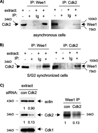
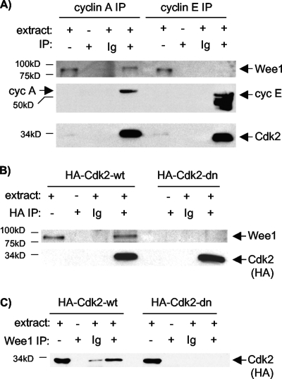
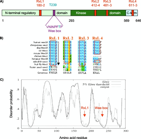
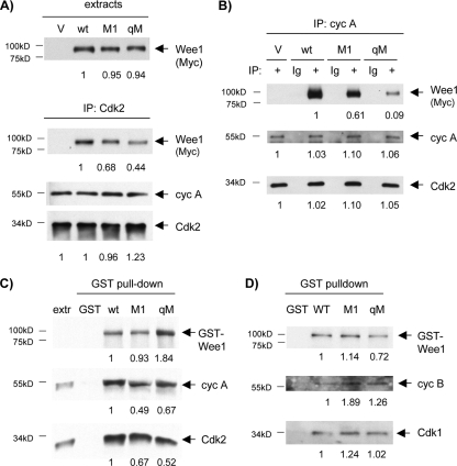
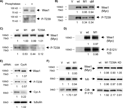
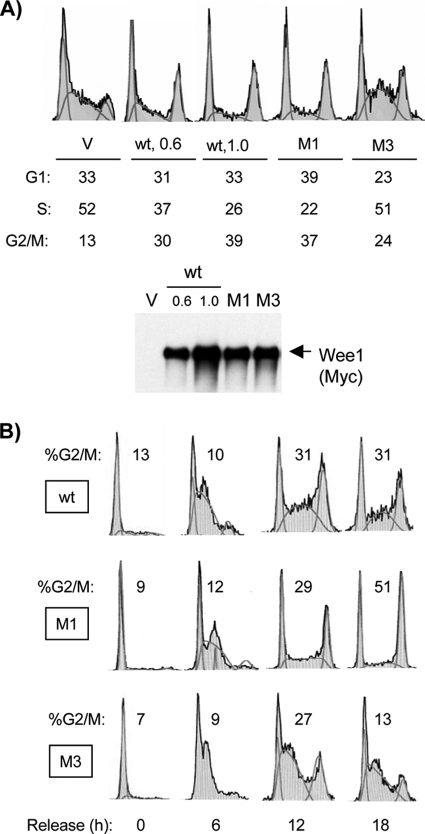
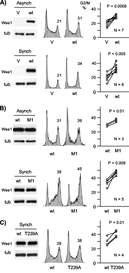
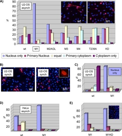
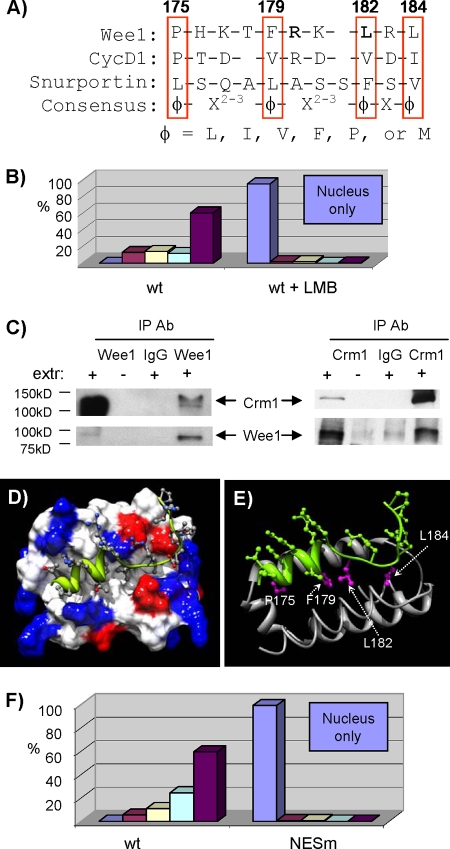
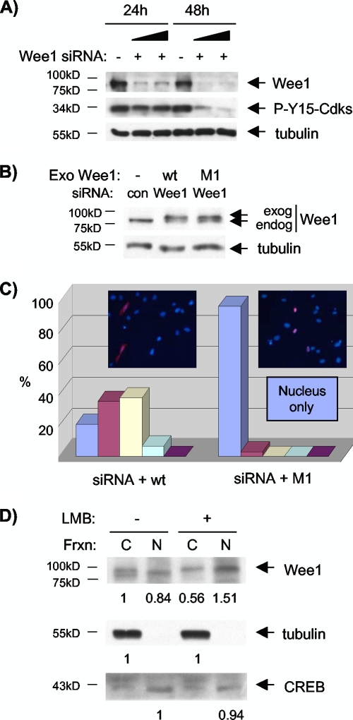
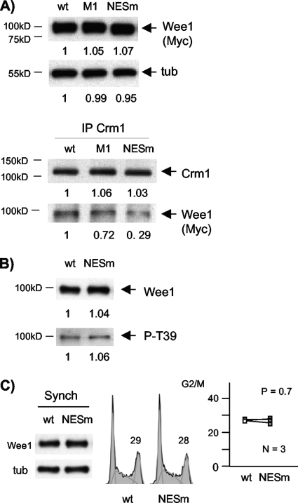
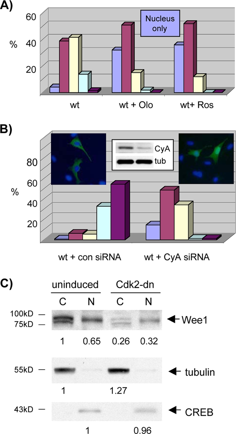
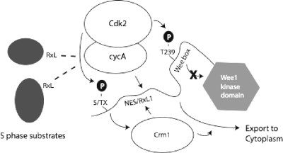
Sophisticated models for the regulation of mitotic entry are lacking for human cells. Inactivating human cyclin A/Cdk2 complexes through diverse approaches delays mitotic entry and promotes inhibitory phosphorylation of Cdk1 on tyrosine 15, a modification performed by Wee1. We show here that cyclin A/Cdk2 complexes physically associate with Wee1 in U2OS cells. Mutation of four conserved RXL cyclin A/Cdk binding motifs (RXL1 to RXL4) in Wee1 diminished stable binding. RXL1 resides within a large regulatory region of Wee1 that is predicted to be intrinsically disordered (residues 1 to 292). Near RXL1 is T239, a site of inhibitory Cdk phosphorylation in Xenopus Wee1 proteins. We found that T239 is phosphorylated in human Wee1 and that this phosphorylation was reduced in an RXL1 mutant. RXL1 and T239 mutants each mediated greater Cdk phosphorylation and G(2)/M inhibition than the wild type, suggesting that cyclin A/Cdk complexes inhibit human Wee1 through these sites. The RXL1 mutant uniquely also displayed increased nuclear localization. RXL1 is embedded within sequences homologous to Crm1-dependent nuclear export signals (NESs). Coimmunoprecipitation showed that Crm1 associated with Wee1. Moreover, treatment with the Crm1 inhibitor leptomycin B or independent mutation of the potential NES (NESm) abolished Wee1 nuclear export. Export was also reduced by Cdk inhibition or cyclin A RNA interference, suggesting that cyclin A/Cdk complexes contribute to Wee1 export. Somewhat surprisingly, NESm did not display increased G(2)/M inhibition. Thus, nuclear export of Wee1 is not essential for mitotic entry though an important functional role remains likely. These studies identify a novel bifunctional regulatory element in Wee1 that mediates cyclin A/Cdk2 association and nuclear export.


Figures

FIG. 1.
FIG. 1.
Wee1 associates with cyclin A/Cdk2 complexes. (A) Wee1 and Cdk2 complexes were immunoprecipitated from lysates of asynchronous U2-OS cells and subjected to immunoblotting (IB) for the opposite protein. Cdk2 was readily detected in Wee1 IPs, and Wee1 was readily detected in Cdk2 IPs. IPs without extract or with nonspecific immunoglobulin G (Ig) served as negative controls. (B) Same experiment as in panel A except that cells were enriched for S and G2 phases, following treatment with HU and release. (C) Cdk2 RNAi confirms association of Wee1 with Cdk2. U2-OS cells were treated with siRNAs directed against Cdk2 or an off-target sequence (control). IB confirmed knockdown of Cdk2 but not Cdk1, thus also confirming the specificity of the respective antibodies (left). Cdk2 RNAi reduced Cdk2 detected in Wee1 IPs (right). Relative band intensity was quantitated, and values are listed below each lane. The numbers below the panels present quantitation of the main bands observed relative to the control (con).
FIG. 2.
FIG. 2.
Lack of Wee1 association with cyclin E or inactive Cdk2 complexes. (A) Cyclin A (cyc A) and cyclin E (cyc E) IPs from asynchronous cells were subjected to IB for Wee1, Cdk2, and the respective cyclins (for the last, the blot was divided, probed separately for each cyclin, and rejoined prior to exposure to film). Note similar recovery of Cdk2 (bottom) in IPs of cyclins A (middle left) and E (middle right) but little detectable Wee1 in the cyclin E IP (top). (B) U2-OS cell clones with inducible expression of HA-Cdk2-wt or HA-Cdk2-dn were induced for 40 h. Protein extracts were immunoprecipitated with anti-HA antibody or nonspecific Ig and subjected to IB for Wee1. Note similar recovery of Cdk2 (bottom). Wee1 associated with Cdk2-wt but not Cdk2-dn. (C) The experiment is the same as that in panel B except that the reciprocal immunoprecipitation used a Wee1 antibody and IB with anti-HA antibody.
FIG. 3.
FIG. 3.
Conserved RXL sequences in Wee1. (A) Primary structure map of human somatic Wee1, with residue numbers at domain boundaries below (black): NRD (light green), kinase domain (dark green), carboxy-terminal domain (light blue), RXL sites (red, with amino acid numbers below), Wee box (single-letter amino acids), and T239 phosphorylation site (aqua). (B) RXL sequences conserved in Wee1 (blue shading). Note that RXL1 and RXL3 are conserved through the fruit fly, and RXL1 is followed by a leucine at position +5, favored in cyclin A binding sequences. The leucine in RXL2 (yellow) is predicted to be involved in packing interactions. (C) Predicted disorder plot of the Wee1 NRD from the DISOPRED server (http://bioinf.cs.ucl.uk/disopred/). The horizontal hatched line marks the 5% default false-positive rate for disordered regions, the solid filter line marks the output from DISOPRED2, and the hatched output line marks the output from a linear support vector machine classifier of lower confidence level. Positions of the RXL1 and Wee box are marked. x axis, Wee1 amino acid sequence; y axis, probability of being disordered.
FIG. 4.
FIG. 4.
Reduced binding of M1 and qM mutants to cyclin A/Cdk2 complexes. (A) U2-OS cells were transiently transfected with empty vector (V), wild-type (wt) Wee1, M1, or qM and enriched in late S and G2 phases by mimosine treatment and release. Protein extracts were subjected to immunoblotting (IB) for the respective Wee1 proteins (top, Myc tag), or immunoprecipitation (IP) for Cdk2 and IB for the respective Wee1 proteins (Myc tag), cyclin A (cyc A), or Cdk2. M1 and qM showed moderately and markedly reduced binding to Cdk2 complexes, respectively. The mean ratio of Cdk2 bound to M1 versus the wild type was 0.72 ± 0.03 (n = 3), and for qM versus the wild type it was 0.40 ± 0.03 (n = 3) (B) The same extracts used in panel A were subjected to cyclin A IP. Nonspecific antibody (Ig) was used for IP as additional negative controls. M1 and qM showed moderately and markedly reduced binding to cyclin A complexes, respectively. The mean ratio of cyclin A bound to M1 versus the wild type was 0.55 ± 0.04 (n = 3), and for qM versus the wild type it was 0.10 ± 0.01 (range, n = 2). (C) GST alone or GST fusion proteins of wild-type Wee1, M1, or qM were incubated with extracts from U2-OS cells enriched for late-S and G2 phases and retrieved on glutathione beads. GST proteins (GST antibody), cyclin A, and Cdk2 were detected by IB. Note that the qM pull-down was somewhat overloaded. M1 and qM showed reduced binding to Cdk2 and cyclin A complexes. Extr: extract alone. The mean ratio of cyclin A bound to GST-M1 versus the wild type was 0.57 ± 0.09 (n = 4), and for GST-qM versus the wild type it was 0.29 ± 0.08 (n = 3). The mean ratio of Cdk2 bound to GST-M1 versus the wild type was 0.80 ± 0.08 (n = 3), and for GST-qM versus the wild type it was 0.29 ± 0.03 (n = 4). (D) GST alone or GST fusion proteins with wild-type Wee1, M1, or qM were used in pull-down assays to assess binding in vitro to cyclin B (cyc B) or Cdk1, detected by IB. Binding of M1 and qM to cyclin B/Cdk1 complexes was not diminished. The numbers below the panels present quantitation of the main bands observed relative to the control.
FIG. 5.
FIG. 5.
Reduced phosphorylation of M1 and qM on T239. G1/S-synchronized U2-OS cells were transfected with the indicated Wee1 proteins, synchronized, and used to prepare extracts from cells enriched in late-S and G2 phases. (A) T239 phosphorylation of Wee1. Anti-P-T239 antibody was used for immunoblotting (IB) of endogenous Wee1 IPs, without or with phosphatase treatment. IB with anti-Wee1 antibody demonstrated a shift in mobility with phosphatase treatment and absence of proteolysis. (B) Reduced T239 phosphorylation in M1 and T239A. Cells expressing wild-type (wt) Wee1, T239A, or M1 were subjected to IB for Wee1 (Myc tag), P-T239, or tubulin (loading control). Note the lack of reactivity of T239A with the P-T239 antibody and reduced reactivity of M1. (C) Reduced T239 phosphorylation in M1 and qM. Extracts from cells expressing wild-type Wee1, M1, or qM were subjected to IB for P-T239. The blot was then stripped and reprobed for Wee1 (Myc tag). The ratios (means ± standard deviations) of P-T239 were as follows: M1 to the wild type, 0.48 ± 0.11 (n = 6), T239A to the wild type, 0.11 ± 0.02 (n = 7), qM to the wild type, 0.40 (range, n = 2). (D) Phosphorylation of S121/123 was unaffected in M1. Extracts from cells transfected with empty vector, the wild type, or M1 were subjected to IB with P-S121/123 or Myc antibody. (E) Cyclin A (CycA) knockdown reduces Wee1 T239 phosphorylation. Extracts of cells transfected with wild-type Wee1 and either scrambled (con) or cyclin A siRNA were subjected to IB for Wee1 (Myc tag), P-T239, cyclin A, or tubulin (loading control). The ratio (mean ± standard deviation) of P-T239 for cyclin A RNAi versus control was 0.52 ± 0.07 (n = 4). (F) M1 and T239A mediate greater Cdk phosphorylated at Y-15 (P-Y15) than the wild type in vivo. Synchronized U2-OS cells were transfected with empty vector (V) and wild-type, M1, T239A, and KD proteins, and cell lysates were prepared from late S/G2-enriched populations. Cdk P-Y15 was assayed by IB. The mean (± standard deviation) ratios of Cdk P-Y15 observed following transfection were as follows: wild type to vector, 1.8 ± 0.2 (n = 4), M1 to wild type, 1.7 ± 0.2 (n = 7), T239A to wild type, 2.2 ± 0.2 (n = 6), KD to wild type, 0.93 ± 0.03 (range, n = 2). The numbers below the panels present quantitation of the main bands observed relative to the control. tub, tubulin.
FIG. 6.
FIG. 6.
Reduced G2/M inhibition by M3 and increased G2/M inhibition by M1. (A) Asynchronous U2-OS cells were transiently transfected with vector (V), wild-type (wt) Wee1 (with two different amounts of plasmid [μg]), M1, and M3; harvested at 40 h; and subjected to flow cytometry (x axis, DNA content; y axis, relative cell number; profiles normalized to the highest peak) or immunoblotting for exogenous Wee1 (Myc tag). The percentages of cells in each cell cycle phase are listed. Note that the M1 G2/M fraction was nearly as high as that of the wild type expressed at substantially higher levels, and the M1 S phase fraction was lower. (B) Cells transfected with the wild type, M1, or M3 were synchronized by mimosine, released for the designated times, and subjected to flow cytometry.
FIG. 7.
FIG. 7.
Increased G2/M arrest mediated by M1 and T239A. (A) Empty vector (V) and wild-type (wt)-expressing plasmids were expressed by transient transfection in asynchronous cells (Asynch) or cells synchronized with mimosine and released for 18 h (Synch). Exogenous Wee1 levels were assayed by immunoblotting (Myc tag), with a tubulin (tub) loading control. Cell cycle position was assayed by flow cytometry from the same plates of cells (center). The percent G2/M content is quantified above that peak. Results from multiple (N) independent experiments are graphed (right), with paired results from the same experiment connected by lines. The wild type yielded consistently more cells in G2/M than vector (means ± standard deviations, 33 ± 6 versus 20 ± 6 for asynchronous cells [P < 0.0008] and 30 ± 2 versus 20 ± 4 for synchronized cells [P < 0.005]). (B) The same experiment as in panel A with transfection of the wild type and M1. M1 consistently yielded higher G2/M levels (46 ± 3 versus 34 ± 6 for asynchronous cells [P < 0.01] and 37 ± 2 versus 30 ± 1 for synchronized cells [P < 0.009]). (C) The same experiment as in panel B except that the wild type and T239A were compared in synchronized cells. T239A yielded consistently higher G2/M levels (40 ± 5 versus 31 ± 6; P < 0.01).
FIG. 8.
FIG. 8.
Increased nuclear localization of M1. (A) Asynchronous (asynch) U2-OS cells were transfected with the respective Wee1 proteins, fixed at 40 h, and subjected to indirect IF staining for Wee1 (Myc tag, red; see inserted images) and DAPI (blue). Cells were scored in a range from exclusively nuclear to exclusively cytoplasmic. Note the dramatically increased nuclear localization of M1. (B) U2-OS cells were transfected with the wild type (wt) or M1, synchronized (synch) by mimosine block and release for 10 h, and stained and scored as described for panel A. M1 was again much more nuclear. Dual-phase contrast/fluorescence microscopy confirmed that the vast majority of M1-expressing cells displayed abundant, broadly spread cytoplasm comparable to wild-type-expressing cells; hence, the stronger nuclear staining of M1 was not secondary to cell rounding (data not shown). (C) Quantitation of Wee1 localization in synchronized U2-OS cells from panel B, scored as in panel A. (D) HeLa cells were transfected with the wild type or M1, fixed at 40 h, and stained and scored as in panel A. (E) M1 and M1KD were expressed in U2-OS cells, and their localizations were scored as per panel A. Each result in this figure is representative of at least two independent experiments.
FIG. 9.
FIG. 9.
RXL1 is embedded within a Crm1-dependent NES. (A) Amino acid sequence alignment of the RXL1 (bold) domain, the consensus Crm1 NES, and the established Crm1-dependent NES from human cyclin D1 (CycD1). Residues matching the consensus are boxed, with the variant residue in a dashed-line box. (B) LMB treatment blocks nuclear export of Wee1. U2-OS cells were transfected with wild-type (wt) Wee1, synchronized at the G1/S border with mimosine, released from the block, and fixed in mid-late S phase. Localization of the exogenous Wee1 was assessed by IF assay using the Myc-tagged antibody. Subcellular distribution was characterized as described in the legend of Fig. 8. Light purple, nucleus only; dark purple, cytoplasm only. (C) Wee1 is physically associated with Crm1. Reciprocal immunoprecipitations (IPs) from an extract (extr) of U2-OS cells synchronized in S phase and subjected to immunoblotting for Wee1 or Crm1. IgG, immunoglobulin G (nonspecific antibody [Ab] control). (D) Model of the Wee1 NES-Crm1 binding interaction, based on Crm1-Snurportin crystal structures. Space-filling was used for Crm1. Blue marks, positive charge; red, negative charge. The Wee1 backbone is shown in green. (E) Ribbon and stick model for the Wee1 NES-Crm1 interaction, with the NES residues highlighted in magenta and labeled by number. (F) Selective mutation of the potential RXL1 Crm1 NES blocks nuclear export. Wild-type Wee1 and a Wee1 mutant in which P175 and F179 were converted to alanine residues (NESm) were expressed by transient transfection in U2-OS cells synchronized in mid-late S phase. Its subcellular localization was assessed by IF as per the legend of Fig. 8.
FIG. 10.
FIG. 10.
Crm1-dependent nuclear export of Wee1 at endogenous levels. (A) U2-OS cells were treated with low and high amounts of Wee1 siRNA for 24 or 48 h. Immunoblotting demonstrated knockdown of endogenous Wee1 and reduction in Cdk Y15 phosphorylation. (B) Expression of exogenous (Exo) proteins at levels similar to endogenous (endog) Wee1, detected by Wee1 immunoblotting. Some signal at the position of endogenous Wee1 in the transfected samples may reflect exogenous protein following proteolysis of the Myc tag. con, control. (C) Nuclear localization of M1 at levels similar to the endogenous protein. The localization of exogenous wild-type (wt) Wee1 and M1 expressed at low levels was assessed by IF and quantified in synchronized cells harvested in mid-to-late S phase, as described in the legend of Fig. 8. (D) LMB treatment inhibits cytoplasmic localization of endogenous Wee1. U2-OS cells were synchronized at the G1/S border, and half of the culture was treated with LMB for the last 4 h. Cells were harvested with the peak in late S/G2. Nuclear (N) and cytoplasmic (C) fractions (Frxn) were subjected to immunoblotting for Wee1. Immunoblotting for tubulin and CREB served as cytoplasmic and nuclear markers, respectively.
FIG. 11.
FIG. 11.
NESm displays reduced association with Crm1 but unperturbed T239 phosphorylation and cell cycle inhibition. (A) Reduced association of NESm with Crm1. Wild-type (wt), M1, and NESm proteins were expressed by transient transfection. Crm1 IPs were assayed by immunoblotting for Wee1 (Myc tag). (B) No change in P-T239 was seen in immunoblotting of the wild type and NESm. (C) No change in G2/M inhibition in NESm. The wild type and NESm were transfected in synchronized cells (Synch) and subjected to flow cytometry, as described in the legend of Fig. 7 (means ± standard deviations, 28 ± 1 versus 28 ± 2; P = 0.7). The numbers below the panels present quantitation of the main bands observed relative to the control. tub, tubulin.
FIG. 12.
FIG. 12.
Wee1 nuclear localization is dependent on cyclin A/Cdk activity. (A) Cdk inhibitor treatment reduces nuclear export of Wee1. U2-OS cells were transfected with wild-type (wt) Wee1, synchronized with mimosine, and released. Cells were left untreated or were treated with olomoucine (Olo) or roscovitine (Ros). Cells were harvested with the peak in mid-S phase and subjected to immunofluorescent staining for Wee1 (Myc tag). Results were quantified as described in the legend of Fig. 8A. (B) Cyclin A RNAi reduces nuclear export of Wee1. U2-OS cells were synchronized with mimosine with transfection of control (con) or Wee1 siRNA (see insert for immunoblot of cyclin A and tubulin control). Cytoplasmic and nuclear fractions were analyzed as described in the legend of Fig. 8A. Each result is representative of two independent experiments. CyA, cyclin A; tub, tubulin. (C) Cdk2-dn induction inhibits nuclear export of Wee1. U2-OS cells were synchronized with mimosine with and without induction of Cdk2-dn. Cells were harvested with the peak in late S/G2. Nuclear (N) and cytoplasmic (C) fractions were subjected to immunoblotting for Wee1. Signal intensity was quantified with ImageJ software and normalized to the Cdk2-dn nuclear fraction. Immunoblotting for tubulin and CREB served as cytoplasmic and nuclear markers, respectively. Each result is representative of two independent experiments. The numbers below the panels present quantitation of the main bands observed relative to the control.
FIG. 13.
FIG. 13.
Model of Wee1 regulation by the NES/RXL1 bifunctional regulatory element. Cyclin A/Cdk complexes are freed from RXL-containing S-phase substrates by accumulation of these Cdk complexes and S-phase progression. The Cdk complexes bind RXL1, which directs phosphorylation of T239 and possibly other sites [(S/T)X] that foster nuclear export of Wee1. T239 phosphorylation abrogates stimulation of kinase activity by the Wee box. Crm1 binds the Wee1 NES and directs nuclear export. cycA, cyclin A.

References

    1. Adams, P. D., W. R. Sellers, S. K. Sharma, A. D. Wu, C. M. Nalin, and W. G. Kaelin. 1996. Identification of a cyclin-Cdk2 recognition motif present in substrates and p21-like cyclin-dependent kinase inhibitors. Mol. Cell. Biol. 16:6623-6633. - PMC - PubMed
    1. Alt, J. R., J. L. Cleveland, M. Hannink, and J. A. Diehl. 2000. Phosphorylation-dependent regulation of cyclin D1 nuclear export and cyclin D1-dependent cellular transformation. Genes Dev. 14:3102-3114. - PMC - PubMed
    1. Baldin, V., and B. Ducommun. 1995. Subcellular localisation of human wee1 kinase is regulated during the cell cycle. J. Cell Sci. 108:2425-2432. - PubMed
    1. Berthet, C., E. Aleem, V. Coppola, L. Tessarollo, and P. Kaldis. 2003. Cdk2 knockout mice are viable. Curr. Biol. 13:1775-1785. - PubMed
    1. Booher, R. N., P. S. Holman, and A. Fattaey. 1997. Human Myt1 is a cell cycle-regulated kinase that inhibits Cdc2 but not Cdk2 activity. J. Biol. Chem. 272:22300-22306. - PubMed

Publication types

MeSH terms