. 2010 Jan;78(1):39-48.
doi: 10.1128/IAI.00691-09. Epub 2009 Oct 26.

Functional characterization of Borrelia spielmanii outer surface proteins that interact with distinct members of the human factor H protein family and with plasminogen

Affiliations

Functional characterization of Borrelia spielmanii outer surface proteins that interact with distinct members of the human factor H protein family and with plasminogen

Annekatrin Seling et al. Infect Immun. 2010 Jan.

Abstract

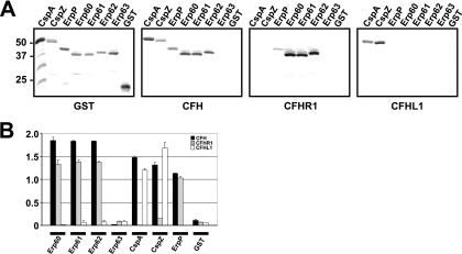
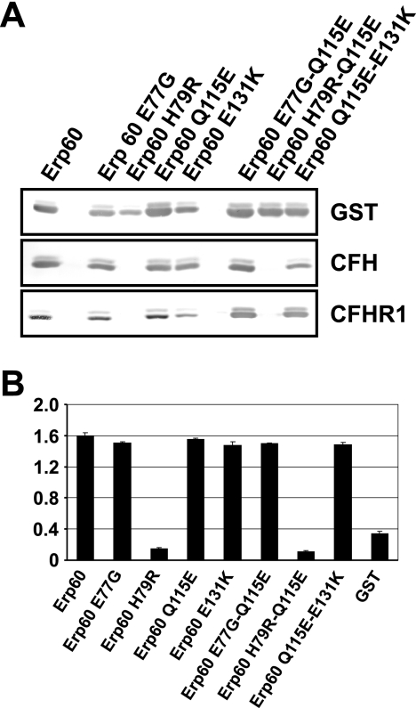
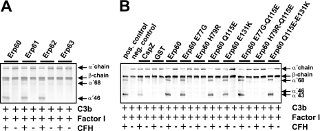
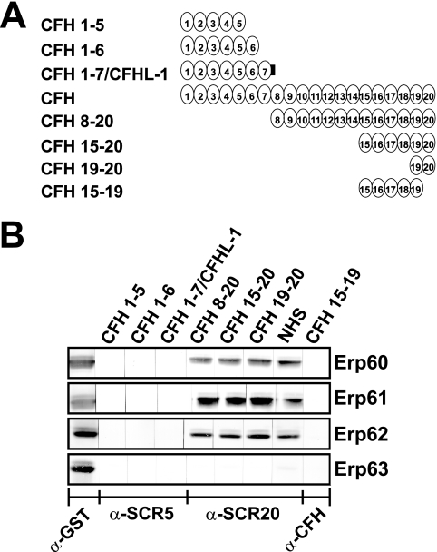
Acquisition of complement regulator factor H (CFH) and factor H-like protein 1 (CFHL1) from human serum enables Borrelia spielmanii, one of the etiological agents of Lyme disease, to evade complement-mediated killing by the human host. Up to three distinct complement regulator-acquiring surface proteins (CRASPs) may be expressed by serum-resistant B. spielmanii, each exhibiting an affinity for CFH and/or CFHL1. Here, we describe the functional characterization of the 15-kDa CRASPs of B. spielmanii, members of the polymorphic Erp (OspE/F-related) protein family, that bind two distinct host complement regulators, CFH and factor H-related protein 1 (CFHR1), but not CFHL1. CFH bound to the B. spielmanii CRASPs maintained cofactor activity for factor I-mediated C3b inactivation. Three naturally occurring alleles of this protein bound CFH and CFHR1 while a fourth natural allele could not. Comparative sequence analysis of these protein alleles identified a single amino acid, histidine-79, as playing a significant role in CFH/CFHR1 binding, with substitution by an arginine completely abrogating ligand binding. The mutation of His-79 to Arg did not inhibit binding of plasminogen, another known ligand of this group of borrelial outer-surface proteins.


Figures

FIG. 1.
FIG. 1.
Serum susceptibility among borrelial isolates. Growth inhibition assays of susceptibility to human serum of B. spielmanii TIsar2 and TIsar3, as well as control strains B. burgdorferi LW2 and B. garinii G1 (27). Spirochetes were incubated in either 50% NHS (filled triangles) or 50% heat-inactivated NHS (open triangles) over a cultivation period of 10 days at 33°C. Color changes were monitored by measurement of the absorbance at 562/630 nm. All experiments were performed three times in which each test was done five times with very similar results. For clarity only data from representative experiments are shown. Error bars represent ± standard deviations (SDs). d, day.
FIG. 2.
FIG. 2.
Deposition of complement components C3, C6, and C5b-9 on the surface of borreliae. Activated complement components deposited on the surface of serum-resistant B. spielmanii isolates TIsar2 and TIsar3, serum-resistant B. burgdorferi LW2, and serum-sensitive B. garinii isolate G1, detected by indirect immunofluorescence microscopy. Spirochetes were incubated with either 25% NHS or hiNHS for 30 min at 37°C with gentle agitation, and bound C3, C6, and C5b-9 were analyzed with specific antibodies against each component and appropriate Alexa Fluor 488-conjugated secondary antibodies. For visualization of the spirochetes in a given microscopic field, the DNA-binding dye DAPI was used. The spirochetes were observed at a magnification of ×100. The data were recorded via a DS-5Mc charge-coupled-device camera (Nikon) mounted on an Olympus CX40 fluorescence microscope. Panels shown are representative of at least 20 microscope fields.
FIG. 3.
FIG. 3.
Identification of factor H and FHL-1 binding proteins expressed among borrelial isolates. Protein extracts (30 μg each) obtained from B. burgdorferi LW2, B. afzelii FEM1-D15, B. garinii G1, and B. spielmanii PC-Eq17, TIsar2, TIsar3, and A14S were separated by 10% Tris-Tricine-SDS-PAGE and transferred to nitrocellulose. The membrane was incubated with purified factor H (2.5 μg/ml), and binding of the proteins was detected with MAb VIG8 specific for SCR 20 of factor H. For detection of FlaB as a control, MAb L41 1C11 was applied. The identified CRASP proteins are indicated on the right, and the mobility of the marker proteins is indicated on the left.
FIG. 4.
FIG. 4.
Alignment of predicted amino acid sequences of the erpA-encoded homologs of B. spielmanii. Amino acid residues identical to those in the sequence of Erp60 from PC-Eq17 are indicated by a dot. Blank spaces in the N terminus of Erp homologs indicate a region for which sequence was not determined due to the cloning procedure used to generate fusion proteins. The lipid-modifiable cysteine residue at position 20 is indicated by an asterisk.
FIG. 5.
FIG. 5.
Analysis of the binding capabilities of CFH, CFHR1, and CFHL1 to recombinant Erp homologs of B. spielmanii. Binding capabilities of CFH, CFHR1, and CFHL1 to recombinant Erp proteins were analyzed by ligand affinity blotting (A). Purified Erp fusion proteins of isolates PC-Eq17, TIsar2, and TIsar3 (500 ng/lane) were subjected to 10% Tris-Tricine-SDS-PAGE and blotted to nitrocellulose membranes. GST fusion proteins were detected by using an anti-goat GST antibody. For detection of CFH and CFHR1 bound to CRASP proteins, membranes were incubated with NHS as a source for CFH or with purified CFHR1. Protein complexes were then visualized using MAb VIG8 or JHD8, respectively. Binding of CFHL1 was detected using MAb B22 specific for the N-terminal region of CFHL1. The CFH/CFHL1-binding CspA and CspZ proteins, the CFH/CFHR1-binding ErpP protein, and purified GST served as controls. Binding was also assayed by ELISA using recombinant Erp60, Erp61, Erp62, Erp63, CspZ, and GST (B). Proteins were immobilized on a microtiter plate and incubated with either CFH, CFHR1, or CFHL1. For detection of protein complexes a polyclonal anti-CFH antiserum was used. All experiments were performed at least three times in which each test was done twice or three times with very similar results, but for clarity data from a representative experiment are shown. Error bars represent standard deviations.
FIG. 6.
FIG. 6.
Identification of amino acid residues within Erp homologs of B. spielmanii responsible for binding of CFH, CFHR1, and CFHL1. Ligand affinity blotting (A) and ELISAs (B) were performed to detect binding of CFH and/or CFHR1 to BsCRASP-3 mutants as described in the legend of Fig. 2.
FIG. 7.
FIG. 7.
Cofactor assay of CFH bound to Erp homologs of B. spielmanii. Erp homologs (A) or Erp mutants (B) immobilized on microtiter plates were used to capture CFH. After sequential addition of C3b and factor I, bound CFH retained cofactor activity by enabling factor I-mediated cleavage of C3b to iC3b. Following incubation, the mixture was separated by SDS-PAGE under reducing conditions and transferred to nitrocellulose, and C3b and degradation products were analyzed using a C3 antiserum (Calbiochem, Darmstadt, Germany). CspZ was used as a positive control for factor I-mediated degradation of C3b. GST and proteins incubated without CFH served as negative controls. The mobility of the α′- and the β-chain of C3 and the cleavage products of the α′ chain the α′-68, α′-46 and α′-43 are indicated.
FIG. 8.
FIG. 8.
Mapping of the domains of CFH responsible for binding to Erp homologs of B. spielmanii. Schematic representation of the CFH protein and CFH deletion constructs (A). Purified recombinant Erps were separated by 10% Tris-Tricine-SDS-PAGE and transferred to nitrocellulose. The membrane strips were incubated with either several constructs of CFH consisting of SCR domains 1 to 5 (CFH1-5), CFH1-6, CFH1-7 (which represents CFHL1), CFH8-20, CFH15-20, CFH19-20, or CFH15-19 or with NHS as a source of CFH (B). Bound proteins were visualized using MAb B22 specific for SCR5 of CFH and CFHL1, MAb VIG8 specific for SCR20 of CFH, or a polyclonal CFH antiserum for detection of bound SCR15-19. GST fusion proteins were detected by using an anti-goat GST antibody. α, anti.
FIG. 9.
FIG. 9.
Erp63 of TIsar3 binds human plasminogen. Results of ELISAs measuring adherence of plasminogen to recombinant Erp63 from B. spielmanii TIsar3, ErpP from B. burgdorferi B31, and bovine serum albumin (BSA). Error bars represent standard deviations. Asterisks indicate ELISA results significantly different from those obtained when BSA was used (P < 0.05).

References

    1. Alitalo, A., T. Meri, T. Chen, H. Lankinen, Z.-Z. Cheng, T. S. Jokiranta, I. J. T. Seppala, P. Lahdenne, P. S. Hefty, D. R. Akins, and S. Meri. 2004. Lysine-dependent multipoint binding of the Borrelia burgdorferi virulence factor outer surface protein E to the C terminus of factor H. J. Immunol. 172:6195-6201. - PubMed
    1. Alitalo, A., T. Meri, P. Comstedt, L. Jeffery, J. Tornberg, T. Strandin, H. Lankinen, S. Bergström, M. Cinco, S. R. Vuppala, D. R. Akins, and S. Meri. 2005. Expression of complement factor H binding immunoevasion proteins in Borrelia garinii isolated from patients with neuroborreliosis. Eur. J. Immunol. 35:3043-3053. - PubMed
    1. Alitalo, A., T. Meri, H. Lankinen, I. Seppala, P. Lahdenne, P. S. Hefty, D. Akins, and S. Meri. 2002. Complement inhibitor factor H binding to Lyme disease spirochetes is mediated by inducible expression of multiple plasmid-encoded outer surface protein E paralogs. J. Immunol. 169:3847-3853. - PubMed
    1. Alitalo, A., T. Meri, L. Ramo, T. S. Jokiranta, T. Heikkila, I. J. T. Seppala, J. Oksi, M. Viljanen, and S. Meri. 2001. Complement evasion by Borrelia burgdorferi: serum-resistant strains promote C3b inactivation. Infect. Immun. 69:3685-3691. - PMC - PubMed
    1. Areschoug, T., M. Stalhammar-Carlemalm, I. Karlsson, and G. Lindahl. 2002. Streptococcal beta protein has separate binding sites for human factor H and IgA-Fc. J. Biol. Chem. 277:12642-12648. - PubMed

Publication types

Associated data