. 2009 Nov 10;106(45):19138-43.
doi: 10.1073/pnas.0908504106. Epub 2009 Oct 26.

Cloning human herpes virus 6A genome into bacterial artificial chromosomes and study of DNA replication intermediates

Affiliations

Cloning human herpes virus 6A genome into bacterial artificial chromosomes and study of DNA replication intermediates

Ronen Borenstein et al. Proc Natl Acad Sci U S A. .

Abstract

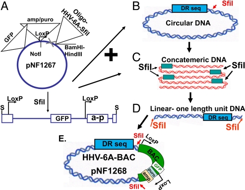
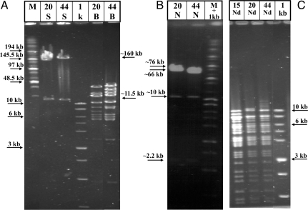
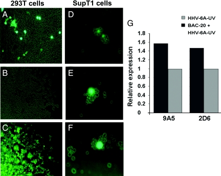
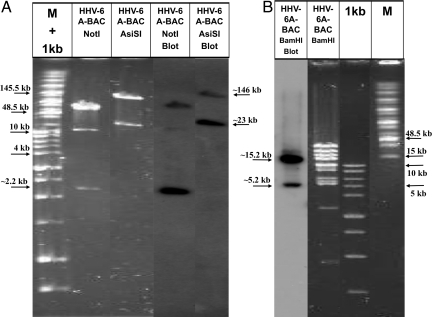
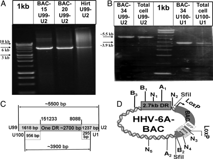
Cloning of large viral genomes into bacterial artificial chromosomes (BACs) facilitates analyses of viral functions and molecular mutagenesis. Previous derivations of viral BACs involved laborious recombinations within infected cells. We describe a single-step production of viral BACs by direct cloning of unit length genomes, derived from circular or head-to-tail concatemeric DNA replication intermediates. The BAC cloning is independent of intracellular recombinations and DNA packaging constraints. We introduced the 160-kb human herpes virus 6A (HHV-6A) genome into BACs by digesting the viral DNA replicative intermediates with the Sfil enzyme that cleaves the viral genome in a single site. The recombinant BACs contained also the puromycin selection gene, GFP, and LoxP sites flanking the BAC sequences. The HHV-6A-BAC vectors were retained stably in puromycin selected 293T cells. In the presence of irradiated helper virus, supplying most likely proteins enhancing gene expression they expressed early and late genes in SupT1 T cells. The method is especially attractive for viruses that replicate inefficiently and for viruses propagated in suspension cells. We have used the fact that the BAC cloning "freezes" the viral DNA replication intermediates to analyze their structure. The results revealed that HHV-6A-BACs contained a single direct repeat (DR) rather than a DR-DR sequence, predicted to arise by circularization of parental genomes with a DR at each terminus. HHV-6A DNA molecules prepared from the infected cells also contained DNA molecules with a single DR. Such forms were not previously described for HHV-6 DNA.


Conflict of interest statement

The authors declare no conflict of interest.

Figures

Fig. 1.
Fig. 1.
The derivation of the HHV-6A-BAC (pNF1268) clone. (A) The pNF1267 clone, containing the amp/puro cassette, the GFP gene, and the oligonucleotide insert was cleaved with the SfiI enzyme. (B) Putative circular replication intermediate, of the HHV-6A genome. The DR sequences of the circularized genome are delineated. (C) Putative head-to-tail concatemers, produced by rolling circle replication. The SfiI site appears once per unit-length viral DNA. (D) SfiI cleavage of the replication intermediates yields unit length genomes with SfiI termini. (E) Ligation of the SfiI-cleaved HHV-6A and the SfiI-cleaved pNF1267, produced the HHV-6A-BAC (pNF1268).
Fig. 2.
Fig. 2.
Restriction enzyme digests of HHV-6A-BAC clones 15, 20, and 44 (pNF1268). The clones were digested with (A) SfiI (S) and BamHI (B), (B) NotI (N), and (C) NdeI (Nd). The digested BACs were analyzed in pulse field gels along with the high molecular weight (M) and 1-kb markers. Some of the marker sizes are noted. Arrows in between A and B point to sizes of fragments of the HHV-6A-BACs. The SfiI fragments of approximately 160 kb, corresponded to the intact HHV-6A genome and the approximate 11.5 kb corresponded to the re-engineered BAC plasmid. Arrows point to the approximate 2.2-kb NotI fragments, containing the amp/puro cassette and the fragment between positions 6,139 bp to 8,332 bp; the approximate 66-kb and 76-kb fragments represent the fragments as expected from the sequence.
Fig. 3.
Fig. 3.
GFP and viral gene expression in 293T and in SupT1-transfected cells. (A) Green fluorescence of 293T transfected cells 72 h p.t. (B) Light photograph of the transfected cells 14 days after puromycin selection. (C) The same as B, but with green fluorescence filter. (D) SupT1 cells electroporated with the pNF1268 and photographed 24 h p.t. (E and F) Cells transfected with pNF1268 and then superinfected with the U.V-inactivated HHV-6A (U1102) virus. The cells were photographed 4 days (E) and 8 days (F) post-infection. (G) Viral gene expression in SupT1 T cells transfected with pNF1268 vector and superinfected with UV light inactivated HHV-6A (U1102). The p41 and gp82–105 proteins reactive with the 9A5 and 2D6 mAb, respectively, were quantified by densitometric analyses. The amounts recovered corrected for loading of the sample relative to the eIF2α “housekeeping” gene. Shown are the average ratios of proteins recovered from cells transfected with pNF1268 and superinfected with HHV-6A-UV compared to the proteins in cells infected solely with the UV-irradiated HHV-6A.
Fig. 4.
Fig. 4.
Restriction enzyme and Southern blot analyses of HHV-6A-BAC with Pac-2-Pac-1 probe. (A and B) Shown are mixtures of the large size marker (M) and the 1 kb marker (lane 1) next to HHV-6A-BAC DNA digested with the restriction enzymes NotI, AsiSI, and BamHI. The Southern blot of the gel was probed with the pac-2-pac-1 probe (A: lane 4 and 5 and B: lane 1). The hybridized bands of approximately 2.2- and 76-kb bands are visible. The band at approximately 10 kb in the blot is most likely non-specific [as apparent from its low intensity (lane 4)].
Fig. 5.
Fig. 5.
PCR analyses of BAC clones, Hirt extracts, total infected cell DNA. (A) PCR products of the BAC-HHV-6A clones 15, 20, and the Hirt preparation of HHV-6A-infected cells DNA employing the denoted primers. (B) PCR of BAC clone 34 and total HHV-6A-infected cells DNA, with the two sets of primers as denoted. (C) A scheme of the PCR results, revealing the presence of a single approximately 2,700-bp DR. The two sets of primers: first set, from U99 up to U2 (positions 149,615 bp sense to 9,325 bp anti-sense yields fragment of ≈5,500 bp). The second set, from U100 up to U1 (positions 150,277 bp sense to 8,383 bp anti-sense yields fragment of ≈3,900 bp). (D) A scheme of the HHV-6A-BAC clone with selected sites of SfiI, NotI (N), AsiSI (A), and the BamHI (B) sites hybridizable to the pac-2-pac-1 probes. The restriction sites are marked with numbers according to their location in the map.

References

    1. Yamanishi K, Mori Y, Pellett PE. In: Fields Virology. 5th Ed. Knipe DM, Howley PM, editors. Lippincott Raven publishers; 2007. pp. 2819–2845.
    1. Ablashi D, et al. Human herpesvirus-6 strain groups: A nomenclature. Arch Virol. 1993;129:363–366. - PubMed
    1. Schirmer EC, Wyatt LS, Yamanishi K, Rodriguez WJ, Frenkel N. Differentiation between two distinct classes of viruses now classified as human herpesvirus 6. Proc Natl Acad Sci USA. 1991;88:5922–5926. - PMC - PubMed
    1. Frenkel N, Borenstein R. Characterization of the lymphotropic Amplicons-6 and Tamplicon-7 vectors derived from HHV-6 and HHV-7. Curr Gene Ther. 2006;6:399–420. - PubMed
    1. Gompels UA, Macaulay HA. Characterization of human telomeric repeat sequences from human herpesvirus 6 and relationship to replication. J Gen Virol. 1995;76:451–458. - PubMed

Publication types

LinkOut - more resources